Education on the eyelid. Is it necessary to remove papillomas on the eyelids? Modern methods of diagnosing the disease
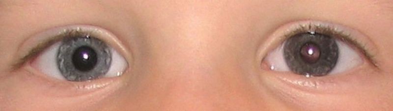
human eye- a kind of camera that has a special hole (pupil) into which rays fall, lenses that focus them, and a retina where the image appears. There are 130,000,000 photosensitive cells inside the eye. When light rays hit them, chemical changes occur, which in turn are converted into a nerve pulse. Through the optic nerve, it enters the part of the brain that is directly responsible for vision. Subsequently, this signal is processed, after which the person sees this or that object.
First, when, by turning the eyelids, it can be seen that they are red, uneven, and bitter, and that the patient complains of burning; this species is called dasites. Secondly, when these symptoms are more severe, and the eyelids form little tubercles, more or less like figs, the disease takes on the name of phycosis, phycosis palpa.
Eyelash disorder that sometimes turns inward and irritates the eyes sharp pains accompanied by inflammation is an evil called trichiasis. When the eyelids stick to each other or to the eye itself, regardless of the cause, this disease is called nodule of the eyelids, and the Greeks - the word consisting of, joins and, eyelid.
eye shape resembles an apple with a convex front part, in the middle is the pupil. The color of the pupil is black due to the black interior of the eye. Through the pupil, light enters the lens, which converts it into an image. Everything is like the film of a camera, there is a layer of light-sensitive cells, or the retina.
Iris is located around the pupil and has the shape of a donut, which can be blue, green, brown. The iris has the ability to change its size. For example: in bright light, the muscles expand, and the pupil narrows, in the dark, everything happens quite the opposite, the pupil expands.
Sometimes the eyelids get so stuck together that we can't open our eyes at all, sometimes this accident happens to just one eye, sometimes to both. sometimes that the eyelid is connected with conjunctivity and more or less strong, in proportion to the number of fibers between which coalition is produced. These kinds of evils come to my eyes when that part or cover that covers has been abused by smallpox, or as a result of " severe inflammation, or "burning, especially if it was done with gunpowder or, in a word, all other expressions that she knows.
Sclera- a sheath that covers eyeball. Proteins are also part of this membrane. From the outside it is transparent, this part of it is called the cornea. The space that exists between the iris and the cornea is filled with "chamber moisture" or liquid. Acts as a lens. The other lens of the eye, the lens, has the ability to change shape. When we look close to ourselves, the lens becomes thicker, but when we look farther away, it becomes thinner.
It is not without example to see children born with this defect and healthy people to write it off on the occasion of fleshy sprouts from one angle or another. Heister saw in his surgery that both things happen. He adds that "he saw the eyelids stuck to the cornea, which is hard to imagine, but in any case with" a rare fact in which it can hardly happen that we treat without losing sight In general, the healing of the eyelid coalition is very vague: one of the cases where it is more difficult to remove the eyelid from above is when the pain is caused by a burn. the best thing to try is to inject, inject into the eyes moisturizing and emollient medicines adapted to keep them always wet and mobile, and to prevent the inflamed parts from sticking to each other.
Eye cancer, what is it?
Causes of eye cancer
The causes of the disease have not been identified to this day, so many doctors believe that every person has a risk of developing eye cancer.
Despite this, the list of risk factors for developing eye cancer is still there:
- genetics - if there are people with this in the family cancer, then the risk of developing eye cancer in offspring increases significantly;
- the nervous state of a person, namely various depressions, problems in the family, depression, contribute to the development of this disease;
- ecology is the main risk factor for all oncological diseases;
- HIV - infection - cases of eye cancer development against the background of HIV infection have been identified;
- secondary eye cancer or metastatic - in case of metastasis of the primary tumor;
- ultraviolet irradiation - people who have overcome the 30-year milestone are recommended to visit tanning salons as little as possible and be under direct sunlight, it is also necessary to protect their eyes from exposure to sunlight with sunglasses;
- Availability age spots on the shell of the eye;
- it is possible a sharp decline immunity against various viral infections.
Types of eye cancer
According to statistics, eye cancer most affects the conjunctiva and eyelid - this occurs in 60% of all cases. Neoplasms that develop inside the eye make up 34%. Orbital cancer is less common, accounting for about 24%. Also, statistics show that malignant neoplasms of the eyes are more common than benign ones.
It is also necessary that the same surgeon, when operating, should try to prevent by appropriate precautions that the eyelids do not reattach. One of good ways to achieve this - set between two very thin linen or a sheet of gold covered with sweet almond oil, they stay there for days until you see him. more afraid of a new coalition.
However, as it often happens that an uncomfortable person cannot suffer anything between his eyelid and his eye, he must then be content with what catches his eye. eye drop plantain water, tuti and saturated sugar and this instillation is often repeated; at the same time the patient will take care to rub gently and move the eyelids himself, spreading them from time to time with his fingers. Therefore, we should expect them to be of reasonable age, especially since this disease is not one of those that are more annoying for a few years.
As you know, tumors of the eyeball are divided into:
- benign;
- malignant.
Benign eye tumors:
- papilloma keratoacanthoma;
- trichoepitheliomas;
- syringoadenomas;
- senile warts on the eyelids;
- benign nevi;
- hemangiomas;
- fibromas;
- neuromas;
- lymphangiomas;
- lipomas;
- fibroids;
- adenomas;
- neurofibromas;
- myxomas;
- gliomas.
Malignant neoplasms include:
- basal cell carcinoma- occurs when sunburn. It is a bump from the bottom of the eye, or on the mucous membrane at the junction of the eyelids. Usually diagnosed in people over 40;
- squamous cell- is an ulcer with pronounced edges, increasing with a certain period of time. If it formed on the edge, there is a danger of going directly into the eye;
- meibomian gland carcinoma (cartilage)- It has yellow, and the shape of this tumor may resemble a chalazion. Usually located in the upper part. characteristic feature The fact that this is precisely this disease is its rapid re-growth at the site of removal. Responds to medications a sharp increase in size;
- , the impetus for the formation of this type of disease is also a thermal burn sunbeams. It mainly affects women and is common among people 40 to 70 years of age. Carries in itself high factor the danger of spreading through the lymph nodes, affects the liver and lungs of a person;
- - is the most common form of intraocular retinal cancer in children. Usually metastasizes to vitreous body and then spreads to the front of the organ;
- eye sarcoma. Develops very quickly and breaks down in a short period of time optic nerve, the mobility of the eyeball decreases, the disease is characterized by strong painful sensations in the affected organ;
- – less dangerous view, because it is not inclined to rapid spread and metastasis. Localization of the tumor on the border area of the epidermis and mucous membrane of the lower eyelid and inner corner of the eye. It manifests itself in the form of a small seal, which increases and forms a crust in its central part. AT individual cases the tumor may grow to a critical large sizes and spreading it to the skin of the cheek and conjunctiva. Metastases can be observed in the cervical and submandibular lymph nodes.
Eye cancer first symptoms and signs of the disease
There are many types of eye cancer, and the symptoms of the disease are directly related to this.
Tumor under Valka's upper eyelid
Valka had a very aggressive tumor that developed 1 month later under the eyelid. On the morning of December 9, she has no problems moving around, not a bad stomach, but still her unhappy look. This frees her and she falls asleep. For Valka there are no diagnostic problems, it's clear, clear, precise. It is trivial to have a wart on the lower eyelid, warts often grow in dogs that take on age. growing outward, it is not aesthetic, but it does not have any irritating effect on the eyes. Under the upper eyelid it is an inflamed gland, it doubles in volume between December 9th and 11th, it irritates the eyeball, but there are no ulcers so far, so we can try to treat with an anti-inflammatory general and topical ointment.
Eye cancer - symptoms of the manifestation of the disease:
- there is a change visual function may be completely lost. This symptom can also appear in other diseases, for example, myopia, but an examination is necessary for an accurate diagnosis;
- an increase in dark spots around the arch of the eye;
- the appearance of sudden pain;
- protrusion of the eye;
- displacement of the eyeball;
- manifestation of strabismus;
- the appearance of frequent flashes before the eyes.
Another variety of tumor is the so-called nevus (mole) on the eye. The appearance is possible from birth, as well as throughout life. If there is an increase in them, you should immediately contact a specialist. These birthmarks can be flat, most often have a convex shape. They may not disturb their owner throughout his life. But there are prerequisites for their degeneration into a malignant tumor.
The operation is not desirable by the vet or by us due to his age. We started treatment, and this morning it has already improved. For the lower eyelid, this is a wart, but for upper eyelid It's not a gland, it's a tumor. For the back, she thinks it's the parrot's beak that's broken, it happens in gay dogs. First she is on cortisone for a few days, then we will see it by improvement or not but for her my bitch still has time ahead of her. This belly is swollen, but she puts it down to greed.
On the this moment neither we nor the veterinarian think about a very aggressive tumor and possible diffusion of metastases to the spine, lungs and water in abdominal cavity. Cortisone adds further discomfort, Valki drinks a lot and pisses under her, especially at night because she can't get up easily and we're too tired to wake up multiple times during the night.

Signs of eye cancer:
- feeling of discomfort in the eyes;
- deterioration of vision;
- eye redness;
- decrease in vitality;
- fatigue and constant malaise;
- loss of appetite and subsequent weight loss for no apparent reason.
As for the signs of eye cancer, they can be diagnosed only on last stage or completely by accident. With malignant neoplasms, peculiar thickenings appear on the outer shell, as well as various papillomas dirty pink. With untimely treatment, the destruction of the eyelid occurs, accompanied by terrible pains. The appearance of various tumors on the eyelid also indicates the development of this disease.
Dog Owner Testimonials
She suffers a lot, can't get up and needs her under her, but until the last moment she follows us in the field, we make her sleep, she is 9 years and 11 months old. Nothing traumatic, banal infection. Testimony of a landowner with adult Liebberg's hypothyroidism.
The only symptom was the visualization of a mobile mass, a greasy appearance, coming from the top of the eye with dirty but not purulent streams. No reduction in visual acuity for both small objects close to the most important in the distance. The behavior of the dog was the same without fatigue or loss of appetite. We thought about training around a small foreign body susceptible to antibiotics. Flows resisting dexamethasone and chloramphenicol, and an augmentation mass, my veterinarian told me to remove this mass.
Important! Each type of cancer starts differently and presents with different symptoms.
Eye cancer in children
Retinoblastoma of the eye in children is the most common form of eye cancer in childhood Every year, retinoblastoma affects 300 small patients in the United States and more more quantities in Russia up to 500 babies. When a disease is detected at an early stage of its development, it can be successfully treated, while almost 90% of young patients manage to restore vision and full health.
At the first appointment, a biopsy was done on the specimen under anesthesia. The verdict of the school laboratory followed; conjunctival mastocytoma. The ophthalmologist sent me to a cancerologist, for whom chemotherapy was the only answer; this case was met twice in the annals of veterinary schools. His weight is always stable.
Care is well supported. She is not tired, continues to play a lot with other dogs, is always very vigilant to guard the territory, and her appetite does not change. Malignant tumors can develop on eyelid A on any other part of the body. The most common type of malignancy is carcinoma a, which comprises 90% of eyelid malignancies. Crayfish and are usually very localized and do not cause training on other parts of the body. In addition, they can increase in depth and reproduce locally.
The development of retinoblastoma occurs not only in the mesh, but also in the nervous tissue, which is concentrated in the back of the eyeball. The disease can be both congenital and after the birth of the baby. The disease is most often diagnosed at the age of 1 to one and a half years.
The main sign of retinoblastoma is a bright spot in the center of his pupil surrounded by a dark rim of the iris.
Cancers are less common but pose a risk of metastasis if they are not treated quickly. Basal or squamous cell carcinoma: slight irritation of the protrusion; no specific symptoms. Carcinoma: a bulge, red, or irritated area on the eyelid; swelling in the eyelid; Croc around the eyes Carcinoma of the sebaceous glands can sometimes be embarrassed by negligence.
: An uneven grain of beauty or a grain of beauty that changes appearance or grows, often, but not all the time, pigmented. What will your doctor be looking for? Inflammation, redness, swelling, ulceration, or distortion of the eyelids. Basal cell carcinoma may appear as a small pearl stained on the lower eyelid. Inflammation in the upper part of the eyelid. Sometimes there is inflammation in the upper part of the eyelid, and if cancer is suspected, the doctor will confirm or deny it by applying it. Prevention Skin cancer is caused by prolonged exposure to the sun, so it's wise to expose it sensibly and wear a pair of glasses that will prevent sun damage to the retina and other sensitive areas of the eye. People with light hair or skin will have a greater predisposition to skin cancer than people with hair called "black" and the skin is more matte.
Retinoblastoma eye symptoms:
- the main symptom of retinoblastoma is the effect of the so-called "cat's eye";
- strabismus, which progresses due to the disease;
- decreased vision;
- the presence of a light spot in the center of the eye.
It is worth noting! The symptoms listed above can also apply to a number of other eye diseases, so these signs alone are not enough to confirm that the baby is suffering from retinoblastoma. Accurate Diagnosis can only be put by a doctor, after a series of examinations.
To prevent cancerous spread, be mindful of slightest symptom: the sooner cancer is reported, the more likely successful treatment. The most commonly used treatment for eyelid cancer is simply surgical removal. It is very important to remove the entire tumor part. Often after removal malignant tumor reconstructive surgery will be performed. The eyelids can be repaired and restored with a simple ophthalmic plastic dressing.
Eyelid cancer can only cause weak visual disturbances, with no real symptoms to warrant routine eye examinations. There is a wide variety of benign tumors that can appear on the eyelids. With some experience, most of them can be identified using biomicroscopy. It is important to distinguish between benign tumors and malignant formations. The main benign tumors in the eyelids are cysts and pigmented tumors. The formation of a cyst in the vast majority of cases is a sign of good quality.
So how do you spot eye cancer?
Diagnosis begins directly with an examination of the eyeball, checking the visual field and its acuity. During the examination, the specialist will definitely interview the patient and collect an anamnesis. Depending on the result, the doctor will prescribe a number of diagnostic tests.
Eye cancer is enough insidious disease, so there is a rather serious question of how to recognize the disease on early stage its development?
On the other hand, a pigmented lesion may be benign or associated with malignancy. benign tumors do not require treatment, but should be regularly monitored. There are 3 main types of malignant tumors of the eyelids.
Basal cell carcinoma Squamous cell carcinoma sebaceous glands. . Basal cell carcinoma is the most common eyelid tumor and accounts for about 85% of all eyelid tumors. There is a connection with sun exposure, and the lower eyelid is the most important.
Treatment is based on complete removal of the tumor, with attention to the removal of all malignant tissue, and depending on the size of the tumor, various methods reconstruction. Squamous cell carcinoma of the eyelid is rare and accounts for about 5% of all eyelid tumors. There is an association with sun exposure, and adults are the most exposed. Squamous cell carcinoma behaves more aggressively than basal cell carcinoma, and with more likely distributed by.
Modern methods disease diagnosis:

- ophthalmoscopy- eye examination. In this case, melanoma is detected very quickly. The doctor looks into the eye with a bright light or lens;
- ultrasound scan- apply sound waves in order to analyze the structure of the eye;
- biopsy- a diagnostic method in which a cell or tissue is taken from the affected area of \u200b\u200bthe eye for the purpose of its further study;
- MRI check (magnetic resonance imaging)- study method internal organs and tissues using the physical phenomenon of magnetic resonance;
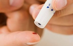
- blood tests to determine the number of leukocytes;
- fluorescein angiography- a method by which you can take a picture of the eye and detect a malignant neoplasm.
To determine the stage of eye cancer, and the spread of the tumor throughout the body will help such diagnostic methods how:
Sebaceous carcinoma is a cancer of the sebaceous glands of the eyelid and accounts for up to 5% of all malignant tumors of the eyelid. This pathology affects mainly the elderly, and women are exposed to greater risk than men. In general, the tumor is located at the level of the upper eyelid. The diagnosis of carcinoma remains difficult: it is often confused with a benign condition such as blepharitis or chalation. As a result, it is often diagnosed at an advanced stage and exhibits aggressive behavior.
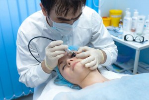
Upper eyelid correction is one of the most common procedures and the most "beneficial" of plastic and cosmetic surgery: a relatively small operation has a big effect, as it widens the field of vision and corrects the sadness of the face.
- Ultrasound of the abdominal cavity;
- blood test for liver function;
- MRI of the brain;
- chest x-ray;
An important point in the conduct and diagnosis is the unmistakable establishment of the type of cancerous tumor. The right treatment strategy depends on this.
Eye Cancer Treatments
In the treatment of eye cancer, it is possible to use different methods, among them are:
- surgical intervention;
- radiation therapy;
- chemotherapy;
- stereotactic radiosurgery;
- eye enucleation;
- brachytherapy;
- alternative treatments.
Informative video: laser eye surgery
eye surgery
At surgical treatment can remove both part of the eyeball, and it completely. These measures are used for advanced stage disease when other methods are not applicable. After the removal of the eye, a person is offered a special prosthesis, it is put in place of the removed eye.
But there is a more gentle operation on the eye, which removes only cancer cells, thereby preserving vision. Depending on the complexity, it has several directions:
- microsurgery (classic excision of the tumor occurs);
- laser (the tumor is removed with a laser);
- radio wave (evaporation of the tumor occurs without contact with the eye).
These directions are modern methods, after which it is possible to save the eye, as well as visual function, since the effect is strictly on the infected areas, without affecting healthy tissues. These therapies are quite expensive, because they are carried out on very expensive equipment.
Radiation therapy for eye cancer
Radiation therapy can be used without or after surgery. It all depends on the degree of tumor damage to the eyes.
Distinguish between internal and external. The method is based on the use of radioactive substances that destroy tumor cells.
Internal radiotherapy uses special grains that are placed in the tissue next to the neoplasm. The residence time is usually 1-2 weeks, then they are removed.
External method of radiotherapy, used in the treatment of eye socket formations. In the process, radioactive rays are directed to the affected area, thereby protecting intact areas.
This method is excellent in the treatment of melanoma. It happens that there are side effects which manifest as dryness and redness of the eyes. Rarely, after such treatment, people can develop cataracts, which are removed. surgically. It is also possible to develop glaucoma and eyelash damage.
Chemotherapy for eyelids
Chemotherapy involves the use of anti-cancer drugs that are given intravenously or taken as a pill. The drugs can also be injected directly into the affected eye, or through spinal cord. Consequently, most of the drug reaches the tumor itself. The chemotherapy treatment cycle is about 3 to 4 weeks.
Side effects after chemotherapy treatment:
- hair loss;
- nausea;
- vomit;
- diarrhea;
- various infections;
- bone marrow suppression;
- fatigue, etc..
Cancer treatment for each patient is selected individually. Selection medical procedures with melanoma of the eye will depend on the location of the tumor and its size. The problem is also radically solved if the eye is red and painful, and intraocular pressure increased. For tumors big size use radiosurgery - irradiation of newly formed tumors. The prognosis depends on the stage of eye cancer at the time of diagnosis and on which parts of the organ are affected.
Stereotactic radiosurgery is a modern trend in the treatment of eye cancer.
Apart from standard method, more modern ones are possible, such as:
- laser infrared radiation;
- laser burning;
- effect on the tumor at low temperatures.
Stereotactic radiosurgery involves treatment with a special metal frame that radiates high energy to the site where the tumor is located. This device is fastened with special screws on the bones of the skull, it is necessary to set the desired direction so that the radiation goes directly to the neoplasm. This method is very painful, so when installing the device, painkillers are used.
Modern technologies have made it possible to create a device that does not need to be fixed on the patient's head. Its powerful doses of energy act only on the affected areas, thereby not affecting other organ systems.
Eye enucleation
Eye enucleation (enucleatio bulbi; lat. enucleare - to extract the nucleus) is an operation to remove the eyeball.
Spend this species operations for various intraocular tumors, both benign and malignant.
Operation contraindications for panophthalmitis - when the disease spreads to the brain and orbital tissues.
Conducting surgery.
The operation is carried out under local anesthesia in adults and under common in childhood. 30 min. before the start of the operation, sodium etaminal 0.1 g and diphenhydramine 0.05 g are prescribed orally, 1 ml is injected under the skin. 1% solution of omnopon. AT conjunctival sac 1% solution of dicaine is instilled, 2 ml is injected retrobulbarno. 2% solution of novocaine, under the conjunctiva of the sclera and along the muscles - 4 ml. 1% novocaine solution.
The eyelid and palpebral fissure are opened with a dilator, grasping the conjunctiva of the sclera at the limbus with tweezers quite widely, incising it with scissors along the circumference of the cornea. The conjunctiva and Tenon's capsule are separated from the sclera along the entire circumference. The end of the muscle hook is inserted under the tendon of the rectus muscles and cut off from the sclera, only the internal (or external) rectus muscle is crossed not at the sclera, but slightly retreating from it, so that a small piece of the tendon remains on the sclera, for which the eyeball is fixed with tweezers. Pulling the eye forward and inserting curved Cooper scissors with closed jaws into the wound from the inside (or outside) behind the eyeball, they grope for the optic nerve; then the scissors are pulled back a little, and open them, moving deeper again and, covering the optic nerve with the brushes, cross it. Consequently, the oblique muscles are crossed at the sclera and the eyeball is removed from the orbit.
Bleeding, which may be present, is stopped with a special swab moistened with a peroxide solution. 3 catgut sutures are applied to the wound of the conjunctiva, a 30% solution of sulfacyl is instilled and a pressure bandage is applied.
Brachytherapy for intraocular tumors
Tumors inside the eyes, quite often secondary malignant neoplasms, that is, caused due to the spread of metastases or cancer cells. However, eye cancer can develop just like the primary tumor. Quite often it is melanoma of the eye or retinoblastoma (cancer of the retina).
Retinoblastoma is most common in childhood; as for melanoma, its development occurs in people of advanced age 60 and older.
There are a lot of methods for the treatment of malignant tumors of the eyes, but one of the most modern is brachytherapy. The main principle of brachytherapy is the installation of radioactive boards, which are installed to further irradiate the tumor of the eye "from the inside", that is, radiotherapy of the eye. Brachytherapy helps reduce cancer.
The first signs of eye cancer can be:
- "blurred picture";
- burning inside the eye;
- redness of the eye;
- tearfulness.
Eye cancer is often asymptomatic, and is diagnosed during a routine examination by an ophthalmologist.
There are 2 stages of brachytherapy for eye cancer:
- stage 1: an operation is performed to install radioactive boards inside the eye. For this operation, use local anesthesia. The operation is carried out in the operating room;
- stage 2: surgery to remove the radioactive board from the eye. The removal of the board takes place a few days after its installation. The term for the board to stay inside the tumor is set only by the attending physician, depending on the size of the tumor and its nature. Throughout the entire period of treatment, the patient must be in the hospital, that is, under the full supervision of a doctor.
Complications during treatment with the use of radioactive boards:
- eye redness;
- intraocular infections;
- retinal disinsertion;
- increased intraocular pressure;
- loss of vision, partial or complete.
In most cases, all of the above complications can be avoided. Brachytherapy in almost all cases avoids enucleation of the eye and stops the growth and spread of a malignant tumor at an early stage of its development.
Alternative Treatments
Complementary therapies include stress relief, meditation, herbal teas and traditional medicine. After chemotherapy, you can do acupuncture. Some methods are safe, as the patient feels better. But, do not forget that all actions should be coordinated only with your doctor.
Who belongs to the risk group?
The reasons why this disease may appear:
- people with fair skin;
- age after 50 years;
- people who spend a lot of time in the sun.
Life expectancy for eye cancer
With a disease such as eye cancer, the prognosis depends entirely on the stage at which it is located, as well as which parts of the eye have been affected. The question is often asked: how long do they live with eye cancer? To answer, experts cite the following statistics:
- upon detection small tumor at an early stage, the survival rate is 85%;
- on the middle - 64%;
- at the latest - 47%.
Disease prevention
Prevention of this group eye ailments consists only in minimizing the causes provoking their occurrence. In addition, each person is required to undergo an annual examination by a qualified ophthalmologist, since deceit cancerous tumors lies in the fact that they almost do not manifest themselves in any way early stages diseases. Of course, such examinations are also mandatory for those who were able to recover from this. dangerous disease threatening not only blindness, but also death.
Xanthelasma of the eyelids is a disease in which a benign flat formation forms on the upper eyelid, which has a yellowish tint and the shape of a raised plaque.
Most often, such a formation is localized near the inner corner of the eye of the upper eyelid. Xanthelasmas can be either single or multiple. In addition, there is such a skin disease as xanthomatosis, in which such benign formations can be located throughout the body, including on the eyelids.
Xanthelasma of the eyelids indicates that the patient's body has abnormalities in fat metabolism, as well as possible deviations in the functionality of the liver.
Thus, this disease is a signal of the body that it occurs serious violations that require urgent treatment.
What are the symptoms of xanthelasma
Xanthelasma of the eyelids is a disease that can be diagnosed quite easily by an experienced specialist based on the results of the examination.
So, consider the signs by which xanthelasma can be determined:
- The formation is small, but at the same time it rises above the surface of the eyelid.
- The shape is slightly flattened and flat.
- Color from lemon to golden yellow.
As for other symptoms, they are practically absent. That is, xanthelasmas do not cause pain and itching.
Xanthelasma of the eyelid is benign neoplasm and has no tendency to degenerate into a malignant tumor.
Formations on the eyelids can increase over time.
Causes
Modern science does not name the exact reasons why eyelid xanthelasmas develop. Doctors adhere to the theory that this neoplasm is one of the varieties of xanthomatosis, and is associated with disorders of cholesterol metabolism in the body.
 According to the results of studies (in which patients with eyelid xanthelasma were studied), people with the following pathologies are at risk of having this disease:
- Overweight, obesity.
- Diabetes.
- Diseases of the liver.
- Pancreatitis.
- Elevated blood cholesterol levels.
xanthomatosis may be hereditary cause occurrence. Therefore, scientists suggest that xanthelasmas can also be inherited. But this theory has no scientific confirmation.
Which specialist to contact?
A disease such as eyelid xanthelasma requires consultation from several specialists at once. These should be:

- Ophthalmologist.
- Endocrinologist.
- Dermatologist.
In addition, the patient will need to take tests that can analyze lipid metabolism.
When diagnosing a disease, doctors should answer the following questions:

- Is the formation on the eyelid exactly a xanthelasma, and not another malignant tumor.
- Can xanthelasma be a consequence of the development of some serious illnesses (diabetes, arteriosclerosis).
- Determining the root causes for the formation of xanthelasma.
 To answer all these questions, the patient will need to undergo a series of examinations. After that, the doctor will prescribe treatment and tell you recommendations about lifestyle.
Xanthelasma may not always have a yellowish tint, in some cases the formation may be reddish in color. In this case, the doctor performs a diascopy during the examination. Its essence lies in the fact that xanthelasma is pressed with a glass slide in order to completely bleed it. After that, you can see the yellow color of the plaque.
How is the treatment carried out
The treatment of xanthelasma of the eyelid is that it is necessary to cure the root cause that caused its development. That is, first of all, it is necessary to restore the disturbed metabolism, or to regulate other disordered functions of the body.
To restore lipid balance and normalize blood cholesterol levels, patients are prescribed lipotropic drugs:

- Clofibrate.
- Parmidin.
- Diosponin.
- Linetol.
- Lipamide.
- Cytamiphene.
AT drug therapy, contributing to the elimination of the root causes of xanthelasma, also include the following pharmacological preparations:
- Vitamin C.
- Calcium pangamat.
- Essentiale.
- A nicotinic acid.
- Pyridoxine.
- Cyanocobalamin.
After eliminating the root cause of xanthelasma, you should not hope that the formation on the eyelid can disappear on its own. Rather, on the contrary, over time, the size of the xanthelasma may increase.
 Therefore, of course, xanthelasma is cosmetic defect with which not every patient agrees to put up with. In this case, doctors may suggest removing the formation using the following methods:
- laser therapy.
- Removal with liquid nitrogen.
- Electrocoagulation.
- surgical method.
Almost all cases of the appearance of xanthelasma on the eyelids are associated with lipid metabolism disorders of varying severity. That is, at its core, xanthelasma is the deposition of cholesterol in the skin.
Therefore, doctors strongly recommend that all patients adhere to such a diet, as well as patients with atherosclerosis. Namely:

- The daily norm should be at least 300 grams of vegetables and 200 grams of fruits. Potatoes are not included in the list of vegetables. At the same time, at least half of the listed vegetables should be consumed raw. Vegetables are very important for the human body, they are rich in all kinds of minerals, vitamins and phytochemical elements, therefore they have positive influence for lipid metabolism.
- Omega-3 fatty acids must be present in the diet. They can be obtained from fatty varieties fish, pumpkin seeds and flax, as well as nuts.
- Water balance. Human body really needs enough liquids. For an adult, the volume of water drunk per day should be from 1.5 to 2 liters. At least 65% of this amount should be ordinary pure water.
- Choose foods that contain fiber. Try to give preference to gray bread or bran. From cereals, choose buckwheat, oats, millet, brown rice and corn. AT daily diet legumes must also be included: peas, lentils, beans, soybeans, chickpeas.
- Instead of animals, it is better to use vegetable fats. These are: corn, olive, sunflower oils.
The list of prohibited products includes:

- Fats of animal origin.
- Bread and bakery products from the highest grade of flour.
- Semolina porridge, white rice, pasta.
- Products with trans fatty acids.
- Alcoholic drinks. This includes even soft drinks.
- Cigarettes and other toxic substances that poison the liver.
- Butter.
 In addition, according to their physical capabilities, patients are encouraged to do daily physical exercises. It can be:
- Jogging in the morning.
- Charger.
- Walking tours.
Combination exercise and a rational diet guarantees the patient a gradual normalization of lipid metabolism in the body. In addition, compliance with these recommendations will avoid the appearance of new xanthelasmas.


