Normal ecg reading. What is an ecg, how to decipher it yourself
An electrocardiogram (ECG) is a record of the electrical activity of heart muscle cells at rest. Professional ECG analysis allows you to assess the functional state of the heart and identify most cardiac pathologies. But this study does not show some of them. In such cases, assigned additional research. So, latent pathology can be detected when taking a cardiogram against the background of a stress test. Holter monitoring is even more informative - taking a round-the-clock cardiogram, as well as echocardiography.
When is an ECG ordered?
The cardiologist issues a referral if the patient has the following primary complaints:
- pain in the heart, back, chest, abdomen, neck;
- swelling in the legs;
- dyspnea;
- fainting;
- interruptions in the work of the heart.
With the sudden appearance of sharp pains in the region of the heart, an ECG should be taken immediately!
Regular removal of a cardiogram is considered mandatory for such diagnosed diseases:
- previous heart attack or stroke;
- hypertension;
- diabetes;
- rheumatism.
Without fail, an ECG is carried out in preparation for operations, pregnancy monitoring, during a medical examination of pilots, drivers, and sailors. The result of the cardiogram is often required when applying for a voucher for sanatorium treatment and issuing permits for active sports activities. For preventive purposes, even in the absence of complaints, it is recommended to take an ECG every year for everyone, especially people over 40 years old. Often this helps to diagnose asymptomatic heart disease.
The heart works tirelessly throughout life. Take care of this amazing organ without waiting for its complaints!
What does the ECG show
Visually, the cardiogram shows a combination of teeth and recessions. The teeth are sequentially designated by the letters P, Q, R, S, T. Analyzing the height, width, depth of these teeth and the duration of the intervals between them, the cardiologist gets an idea about the state of different parts of the heart muscle. So, the first P wave contains information about the work of the atria. The next 3 teeth represent the process of excitation of the ventricles. After the T wave, there is a period of relaxation of the heart.

An example of an ECG fragment with a normal sinus rhythm
The cardiogram allows you to determine:
- heart rate (HR);
- heart rate;
- various types of arrhythmias;
- various types of conduction blocks;
- myocardial infarction;
- ischemic and cardiodystrophic changes;
- Wolf-Parkinson-White syndrome (WPW);
- ventricular hypertrophy;
- the position of the electrical axis of the heart (EOS).
Diagnostic value of ECG parameters
heart rate
The heart of an adult human normally contracts from 60 to 90 times per minute. At a lower value, bradycardia is determined, and at a higher value, tachycardia, which is not necessarily a pathology. So, significant bradycardia is characteristic of trained athletes, especially runners and skiers, and transient tachycardia is quite normal with mental experiences.

In healthy adults, the pulse rate corresponds to the heart rate and is equal to 60 -90 for 1 minute
Heartbeat
Normal heartbeat called regular sinus, i.e., generated in the sinus node of the heart. Non-sinus generation is pathological, and irregularity indicates one of the types of arrhythmia.
During the ECG, the patient is asked to hold their breath in order to identify a possible pathological non-respiratory arrhythmia. A serious problem is atrial fibrillation (atrial fibrillation). With it, the generation of cardiac impulses occurs not in the sinus node, but in the cells of the atria. As a result, the atria and ventricles contract randomly. This contributes to thrombosis and creates a real threat of heart attack and stroke. To prevent them, lifelong antiarrhythmic and antithrombotic therapy is prescribed.
Atrial fibrillation - quite frequent illness in old age. It may be asymptomatic, but pose a real threat to health and life. Follow your heart!
Arrhythmia also includes extrasystole. An extrasystole is an abnormal contraction of the heart muscle under the influence of an excess electrical impulse that does not come from sinus node. There are atrial, ventricular and atrioventricular extrasystoles. What types of extrasystoles require intervention? Single functional extrasystoles (usually atrial) often occur with a healthy heart against the background of stress or excessive physical exertion. Potentially dangerous include group and frequent ventricular extrasystoles.
blockades
Atrioventricular (A-V) blockade is a violation of the conduction of electrical impulses from the atria to the ventricles. As a result, they contract out of sync. A-V block usually requires treatment and, in severe cases, a pacemaker.
Impaired conduction within the myocardium is called bundle branch block. It can be localized on the left or right leg or both together and be partial or complete. With this pathology, conservative treatment is indicated.
Sinoatrial blockade is a conduction defect from the sinus node to the myocardium. This type of blockade occurs with other heart diseases or with an overdose. medicines. Requires conservative treatment.
myocardial infarction
Sometimes an ECG reveals a myocardial infarction - necrosis of a section of the heart muscle due to a cessation of its blood circulation. The cause may be large atherosclerotic plaques or a sharp vasospasm. The type of infarction is distinguished by the degree of damage - small-focal (not Q-infarction) and extensive (transmural, Q-infarction) types, as well as localization. Detection of signs of a heart attack suggests urgent hospitalization of the patient.

ECG for myocardial infarction
The detection of scars on the cardiogram indicates a past myocardial infarction, possibly painless and unnoticed by the patient.
Ischemic and dystrophic changes
Ischemia of the heart is called oxygen starvation of its various parts due to insufficient blood supply. The detection of such a pathology requires the appointment of anti-ischemic drugs.
Dystrophic refers to metabolic disorders in the myocardium that are not associated with circulatory disorders.
Wolff-Parkinson-White syndrome
it congenital disease, which consists in the existence of abnormal conduction pathways in the myocardium. If this pathology causes arrhythmic attacks, then treatment is necessary, and in severe cases, surgical intervention.
Hypertrophy of the ventricles - an increase in size or thickening of the wall. Most often, hypertrophy is a consequence of heart defects, hypertension, and pulmonary diseases. The position of the EOS has no independent diagnostic value either. In particular, with hypertension, a horizontal position or deviation to the left is determined. The composition also matters. In thin people, as a rule, the position of the EOS is vertical.
Features of the ECG in children
For children under the age of one year, tachycardia up to 140 beats per minute, fluctuations in heart rate when taking an ECG, are considered normal. incomplete blockade right leg of the bundle of His, vertical EOS. At the age of 6 years, a heart rate of up to 128 beats per minute is acceptable. Respiratory arrhythmia is typical for the age of 6 to 15 years.
The method of electrocardiography is a simple and painless method of non-invasive diagnostics of the work of internal organs, which does not bring discomfort and does not directly affect the body. However, it is also an extremely informative examination, which is what has made it so popular for a long time. Only, unlike ultrasound studies, a cardiogram does not emit any waves, but only reads information, therefore, in order to find out what the ECG actually shows, it is necessary to turn to the principle of operation of the device itself. The electrocardiograph has a system of sensors that are attached to certain places on the patient's body and record the information received from there. All these highly sensitive mechanisms are able to capture the signals of electrical impulses produced by the work of the heart, and transform them into a curve, each tooth of which has its own special meaning. Thanks to this, doctors have the opportunity to quickly and easily identify various possible pathologies and abnormalities in the work of the heart and cardiovascular system, and even find out which diseases led to this. The simplicity and accessibility of this procedure allows it to be carried out quite often as a preventive diagnosis, and also as the very first and quick examination, which is performed if heart disease is suspected.
Although this procedure examination has been used for the diagnosis of cardiovascular diseases for many years, it remains relevant to the present, due to its availability for patients and its effectiveness. The results obtained during the examination are an accurate reflection of the process occurring inside the human myocardium.
What does a heart cardiogram show?
The cardiogram reflects the rhythm of the heart and its impulses that are produced during work, and also captures the pulse, conductivity and the time it takes for the body to fill with blood. All this makes it possible to draw up a fairly complete clinical picture of the electrical activity of the myocardium and the general condition of the heart. All information transmitted from the sensors is recorded on the tape and compared with the results that should be normal for a person. If pathologies are present, they will necessarily be reflected on the cardiogram in the form of deviations of the main teeth of the curve. By what kind of teeth they are and how exactly they differ from the norm, the doctor can make a conclusion about the diagnosis of the patient, since each pathology is characterized by a certain set of deviations.
Thus, the electrocardiogram allows you to determine how fast the ventricles of the heart fill, identify myocardial problems and notice heart rhythm disturbances and the frequency of its contractions. The method makes it possible to learn about the state muscle tissue due to the fact that the injured myocardium transmits impulses differently than healthy muscles. These changes are able to detect highly sensitive sensors on the patient's skin.
Often, in addition to the presence of pathology, the doctor can determine the type of damage and its location on the heart. A qualified cardiologist is able to identify deviations from the norm by the angles of inclination of the cardiogram teeth, without confusing them with normal variants, and make a diagnosis.
It would not be superfluous to take the results of previous electrocardiographic studies with you to an appointment with a cardiologist so that the doctor can determine the dynamics of the state of the heart and cardiovascular system, as well as track changes in rhythm, calculate whether the heart rate has increased, and whether any pathologies have appeared. All this will help to timely diagnose the development of diseases that can cause diseases such as myocardial infarction and will help to start treatment in a timely manner.
Diseases of the cardiovascular system, which can be determined by ECG
- Arrhythmia. Arrhythmia is characterized by a violation in the formation of an impulse and its progress through the muscle layer. At the same time, rhythm failure is often noted, the time intervals between R - R increase when the rhythm changes, and slight fluctuations in P - Q and Q - T become noticeable;
- Angina. This disease leads to pain in the heart. the cardiogram in this pathology shows a change in the amplitude of the T wave and depression of the S-T segment, which can be seen in certain parts of the curve;
- Tachycardia. With this pathology, there is a significant increase in contractions of the heart muscle. On the ECG, tachycardia is determined by a decrease in the intervals between segments, an increase in the rhythm, as well as a shift in the RS-T part by a small distance;
- Bradycardia. This disease is characterized by a reduced frequency of myocardial contractions. The ECG picture with such a pathology differs from the norm only by a decrease in rhythm, an increasing interval between segments and a slight change in the amplitude of the teeth;
- Hypertrophy of the heart. This pathology is determined by an overload of the ventricles or atria and manifests itself on the cardiogram in the form of an increased amplitude of the R wave, impaired tissue conductivity, as well as an increase in time intervals for an enlarged myocardial area and a change in the electrical position of the heart itself;
- Aneurysm. The aneurysm is manifested by finding a QS wave at the high R site and an elevated RS–T segment at the Q site;
- Extrasystole. With this disease, a rhythm disturbance appears, the ECG shows a large pause after extrasystoles, QRS deformation, altered extrasystoles and the absence of a P (e) wave;
- Pulmonary embolism. For similar pathology characteristic oxygen deficiency of muscle tissue, hypertension of the vessels of the pulmonary circulation and an increase in the right heart, overload of the right ventricle and supraventricular tachyarrhythmias;
- Myocardial infarction. A heart attack can be identified by the absence of the R wave, the rise of the S-T segment and the negative T wave. During the acute stage on electrocardiography, the S-T segment is located above the isoline, and the T wave is not differentiated. The subacute stage is characterized by the descent of the S-T region and the appearance of a negative T. At the stage of infarction scarring, the ECG shows that the S-T segment is isoelectric, T is negative, and the Q wave is clearly visible.
Diseases that are difficult to diagnose using an ECG
In most cases, ECG does not allow diagnosing diseases such as malignant and benign neoplasms in the heart area, defective state of blood vessels and birth defects heart, as well as disturbances in the dynamics of the blood. However, in most cases, due to its location, tumors in different departments hearts affect the work of the muscle and cause disturbances in intracardiac dynamics, which are diagnosed by ECG as valvular defects of the organ. Therefore, in the case when a cardiologist reveals such disorders during the diagnosis process as hypertrophy of the heart, uneven or irregular rhythm, as well as heart failure, he can additionally prescribe echocardiography after the ECG, which will help determine whether there are neoplasms in the heart or the patient has another disease .
The problem with the ECG is that the initial stages of some diseases, as well as certain types of pathologies, are poorly visible on the cardiogram. This is due to the fact that the time of the procedure is not enough to make a full examination and examine the patient's heart in different situations. As a solution to this problem based on electrocardiography, there is a diagnostic method in which the patient must walk with a device that measures heart health for a day or even more.
Congenital heart defects include a whole group of diseases that lead to pathologies in the work of the myocardium. However, during echocardiography, such heart defects are usually identified as signs of specific syndromes, such as hypoxia or heart failure, due to which it is difficult to identify the underlying cause of the disease.
Also, a great difficulty for diagnosis using an ECG is the fact that some pathologies have similar disorders and deviations, which are noted by the cardiogram. In this case, it is necessary to consult an experienced cardiologist, who, according to the results obtained, will be able to give more accurate diagnosis or send it for further testing.
Another problem of electrocardiography is that in most cases the procedure occurs when the patient is at rest, while for ordinary life the absence of physical activity and psycho-emotional arousal is absolutely atypical for most people. Thus, in some cases, with an ECG without additional voltage, an inaccurate clinical picture is obtained, which can affect the final results of the diagnosis, since in most cases symptoms and pathologies do not appear in a calm state. That is why, for maximum efficiency of the study, the electrocardiography procedure can occur with minor patient loads or immediately after them. This provides more accurate information about the state of the heart and the presence of possible pathologies.
Definition of myocardial infarction using a cardiogram
Myocardial infarction is divided into several stages. The first one is acute period, in which part of the muscle tissue dies, while on the cardiogram at this stage of the disease, the excitation vector disappears in those parts of the heart where myocardial damage occurred. Also on the ECG it becomes clear that there is no R wave and Q appears, which normally should not be in the leads. At the same time, the location of the S-T region also changes and the appearance of the T wave is diagnosed. After the acute stage, comes subacute period, at which the indicators of the T and R teeth gradually begin to return to normal. At the stage of scarring, the heart gradually adapts to tissue damage and continues its work, while the scar left after a heart attack is clearly visible on the cardiogram.
Determination of ischemia using ECG
Ischemic heart disease is characterized by reduced blood supply to the myocardium and other tissues of the heart, resulting in a lack of oxygen and gradual muscle damage and atrophy. Too long oxygen deficiency, often characteristic of the advanced stage of ischemia, can subsequently lead to the formation of myocardial infarction.
ECG is not the most good method, which allows you to identify ischemia, since this procedure is performed at rest, in which it is quite difficult to diagnose the location of the affected area. Also, there are certain areas on the heart that are not available for examination by electrocardiography and are not tested, therefore, if a pathological process occurs in them, this will not be noticeable on the ECG, or the data obtained may subsequently be interpreted by the doctor incorrectly.
On the ECG, coronary heart disease is manifested, first of all, by disturbances in the amplitude and shape of the T wave. This is due to reduced impulse conduction.
What can an electrocardiogram tell?
An electrocardiogram (ECG) is a record of the electrical activity of heart muscle cells at rest. Professional ECG analysis allows you to assess the functional state of the heart and identify most cardiac pathologies. But this study does not show some of them. In such cases, additional studies are prescribed. So, latent pathology can be detected when taking a cardiogram against the background of a stress test. Holter monitoring is even more informative - taking a round-the-clock cardiogram, as well as echocardiography.
When is an ECG ordered?
The cardiologist issues a referral if the patient has the following primary complaints:
- pain in the heart, back, chest, abdomen, neck;
- swelling in the legs;
- dyspnea;
- fainting;
- interruptions in the work of the heart.
Regular removal of a cardiogram is considered mandatory for such diagnosed diseases:
- previous heart attack or stroke;
- hypertension;
- diabetes;
- rheumatism.
Without fail, an ECG is carried out in preparation for operations, pregnancy monitoring, during a medical examination of pilots, drivers, and sailors. The result of the cardiogram is often required when applying for a voucher for sanatorium treatment and issuing permits for active sports activities. For preventive purposes, even in the absence of complaints, it is recommended to take an ECG every year for everyone, especially people over 40 years old. Often this helps to diagnose asymptomatic heart disease.
The heart works tirelessly throughout life. Take care of this amazing organ without waiting for its complaints!
What does the ECG show
Visually, the cardiogram shows a combination of teeth and recessions. The teeth are sequentially designated by the letters P, Q, R, S, T. Analyzing the height, width, depth of these teeth and the duration of the intervals between them, the cardiologist gets an idea about the state of different parts of the heart muscle. So, the first P wave contains information about the work of the atria. The next 3 teeth represent the process of excitation of the ventricles. After the T wave, there is a period of relaxation of the heart.
The cardiogram allows you to determine:
- heart rate (HR);
- heart rate;
- various types of arrhythmias;
- various types of conduction blocks;
- myocardial infarction;
- ischemic and cardiodystrophic changes;
- Wolf-Parkinson-White syndrome (WPW);
- ventricular hypertrophy;
- the position of the electrical axis of the heart (EOS).
Diagnostic value of ECG parameters
The heart of an adult human normally contracts from 60 to 90 times per minute. At a lower value, bradycardia is determined, and at a higher value, tachycardia, which is not necessarily a pathology. So, significant bradycardia is characteristic of trained athletes, especially runners and skiers, and transient tachycardia is quite normal with mental experiences.
Heartbeat
A normal heart rhythm is called regular sinus, that is, generated in the sinus node of the heart. Non-sinus generation is pathological, and irregularity indicates one of the types of arrhythmia.
During the ECG, the patient is asked to hold their breath in order to identify a possible pathological non-respiratory arrhythmia. A serious problem is atrial fibrillation (atrial fibrillation). With it, the generation of cardiac impulses occurs not in the sinus node, but in the cells of the atria. As a result, the atria and ventricles contract randomly. This contributes to thrombosis and creates a real threat of heart attack and stroke. To prevent them, lifelong antiarrhythmic and antithrombotic therapy is prescribed.
Atrial fibrillation is a fairly common disease in old age. It may be asymptomatic, but pose a real threat to health and life. Follow your heart!
Arrhythmia also includes extrasystole. An extrasystole is an abnormal contraction of the heart muscle under the influence of an excess electrical impulse that does not originate from the sinus node. There are atrial, ventricular and atrioventricular extrasystoles. What types of extrasystoles require intervention? Single functional extrasystoles (usually atrial) often occur with a healthy heart against the background of stress or excessive physical exertion. Potentially dangerous include group and frequent ventricular extrasystoles.
blockades
Atrioventricular (A-V) blockade is a violation of the conduction of electrical impulses from the atria to the ventricles. As a result, they contract out of sync. A-V block usually requires treatment and, in severe cases, a pacemaker.
Impaired conduction within the myocardium is called bundle branch block. It can be localized on the left or right leg or both together and be partial or complete. With this pathology, conservative treatment is indicated.
Sinoatrial blockade is a conduction defect from the sinus node to the myocardium. This type of blockade occurs with other heart diseases or with an overdose of drugs. Requires conservative treatment.
myocardial infarction
Sometimes an ECG reveals a myocardial infarction - necrosis of a section of the heart muscle due to a cessation of its blood circulation. The cause may be large atherosclerotic plaques or a sharp vasospasm. The type of infarction is distinguished by the degree of damage - small-focal (not Q-infarction) and extensive (transmural, Q-infarction) types, as well as localization. Detection of signs of a heart attack suggests urgent hospitalization of the patient.
The detection of scars on the cardiogram indicates a past myocardial infarction, possibly painless and unnoticed by the patient.
Ischemic and dystrophic changes
Ischemia of the heart is called oxygen starvation of its various parts due to insufficient blood supply. The detection of such a pathology requires the appointment of anti-ischemic drugs.
Dystrophic refers to metabolic disorders in the myocardium that are not associated with circulatory disorders.
Wolff-Parkinson-White syndrome
This is a congenital disease, consisting in the existence of abnormal conduction pathways in the myocardium. If this pathology causes arrhythmic attacks, then treatment is necessary, and in severe cases, surgical intervention.
Hypertrophy of the ventricles - an increase in size or thickening of the wall. Most often, hypertrophy is a consequence of heart defects, hypertension, and pulmonary diseases. The position of the EOS has no independent diagnostic value either. In particular, with hypertension, a horizontal position or deviation to the left is determined. The composition also matters. In thin people, as a rule, the position of the EOS is vertical.
Features of the ECG in children
For children under the age of one year, tachycardia up to 140 beats per minute, fluctuations in heart rate when taking an ECG, incomplete blockade of the right leg of the His bundle, vertical EOS are considered normal. At the age of 6 years, a heart rate of up to 128 beats per minute is acceptable. Respiratory arrhythmia is typical for the age of 6 to 15 years.
What does electrocardiography (ECG) show?
Conditions of myocardial infarction, angina pectoris, atherosclerosis, myocardiopathy, rheumatic heart disease, arrhythmias of various origins, hypertension - all these cardiac diseases occur in people over the age of forty.
Heart disease occurs due to the negative impact on the human body of certain hereditary factors, chronic overstrain (emotional or physical), physical trauma, stress or neuroses.
I recently read an article that talks about Monastic tea for the treatment of heart disease. With the help of this tea, you can FOREVER cure arrhythmia, heart failure, atherosclerosis, coronary heart disease, myocardial infarction and many other diseases of the heart and blood vessels at home.
I was not used to trusting any information, but I decided to check and ordered a bag. I noticed changes within a week. constant pain and the tingling in my heart that had tormented me before - receded, and after 2 weeks disappeared completely. Try it and you, and if anyone is interested, then below is a link to the article.
Also, common reasons for the development of one or another cardiovascular pathology may become: wrong image life, irrational nutrition, bad habits, sleep and wakefulness disturbances.
But today, we would like to talk about that. In today's publication, we propose to pay attention to the electrocardiography (ECG) procedure, with the help of which doctors are able to detect these pathologies in a timely manner.
What is this diagnostic technique? What does a cardiogram show to doctors? How informative and safe is the procedure in question?
Maybe, instead of a banal cardiogram (ECG), it is better to conduct an ultrasound examination (ultrasound) of the heart? Let's figure it out.
What deviations in the work of the body can be fixed?
First of all, it should be noted that the procedure of electrocardiography (ECG) is deservedly recognized as the main diagnostic technique for the timely detection of pathologies of the heart (the entire cardiovascular system). The procedure is widely used in modern cardiology practice.
The muscular structure of the human heart functions under the constant control of the so-called pacemaker, which originates in the heart itself. At the same time, its own pacemaker generates electrical impulses that are transmitted through the conduction system of the heart to its various departments.
Muscular structure of the heart
On any version of the cardiogram (ECG), it is precisely these electrical impulses that are recorded and recorded, which make it possible to judge the functioning of the organ.
In other words, we can say that the ECG captures and records the peculiar language of the heart muscle.
According to the resulting deviations of specific teeth on the cardiogram (recall, these are the P, Q, R, S and T teeth), doctors get the opportunity to judge what pathology underlies the unpleasant symptoms felt by the patient.
For the treatment of cardiovascular diseases, Elena Malysheva recommends a new method based on Monastic tea.
It contains 8 useful medicinal plants, which are extremely effective in the treatment and prevention of arrhythmia, heart failure, atherosclerosis, coronary artery disease, myocardial infarction, and many other diseases. In this case, only natural ingredients are used, no chemicals and hormones!
With the help of various ECG options, doctors can recognize the following diseases hearts:
Hypertrophy of various parts of the heart muscle.
The problem may occur with hemodynamic disorders vascular bed, which provokes an overload of various cardiac departments. Even a classic ECG allows you to fix several basic signs of cardiac hypertrophy.
These can be: signs of an increase in the behavior of impulses, changes in the amplitude of various teeth, signs of ischemia of the subendocardial cardiac sections, deviation of the electrical cardiac axis.
Angina pectoris, ischemic heart disease.
Having studied the methods of Elena Malysheva in the treatment of HEART DISEASE, as well as the restoration and cleaning of VESSELS, we decided to bring it to your attention.
This disease, we recall, gives a lot of trouble to a person, since it is manifested by attacks of anginal pain that can last from insignificant seconds to half an hour.
Signs of this disease on the ECG can be recorded: as changes in the QRS complexes, as a state of depression of the S-T segment, changes in the T wave.
Arrhythmias of various types.
Such pathologies of the heart muscle are incredibly diverse, they are characterized by numerous changes in the rhythm of heart contractions. On electrocardiography, such violations are manifested: by the frequency of changes in the R-R intervals, by fluctuations in the P-Q and Q-T indicators.
In addition, with the help of electrocardiography, it is often possible to fix: signs of the presence of a heart aneurysm, the development of extrasystole, the occurrence of an inflammatory process in the myocardium (myocarditis, endocarditis), the development of acute conditions of myocardial infarction or heart failure.
Do the results of different ECG methods differ?
It is no secret to anyone that electrocardiography in different situations can be carried out in different ways, or rather, doctors can use various methods ECG research.
It is quite clear that the data of various variants of an electrocardiographic study may differ somewhat.
The most common electrocardiographic studies can be considered:
Intraesophageal electrocardiography procedure.
The technique consists in placing an active electrode in the lumen of the esophagus.
This procedure allows more accurate assessment of atrial electrical activity, as well as the functioning of the atrioventricular node.
The technique is of greatest value for fixing certain heart blocks.
Vectorcardiography procedure. This technique allows you to register changes in the electrical vector of the functioning of the heart muscle.
Information can be presented in the form of a special projection of three-dimensional figures on the plane of assignments.
Electrocardiographic tests with a load.
This procedure may also be called bicycle ergometry. It is most expedient to conduct such a study to detect coronary disease of the heart muscle.
This is due to the fact that angina attacks usually occur precisely at the moment of the patient's physical exertion, and at rest the cardiogram may remain within the normal range.
Holter monitoring procedure.
The procedure is commonly referred to as 24-hour Holter electrocardiography monitoring.
The essence of the technique lies in the fact that the sensors fixed on the human body record the performance of the heart muscle during the day or even more.
It is most appropriate to carry out such a procedure when unpleasant symptoms heart disease are transient.
What diseases can be diagnosed during the study?
It should be said that various options for electrocardiography of the heart can be used not only as a primary diagnosis, which makes it possible to fix the initial stages of a cardiac disease.
Often, electrocardiographic studies various types can be carried out for the purpose of monitoring and controlling an already existing cardiac pathology.
So such studies can be prescribed to patients with the following pathologies:
- patients with previous myocardial infarction;
- people suffering from various forms cardiac ischemia;
- patients with infectious diseases of the heart muscle - pericarditis, endocarditis;
- patients suffering from cardiosclerosis;
- people with hypertension or hypotension;
- patients with vegetovascular dystonia etc.
And, of course, this study of the heart often allows you to answer questions - why do patients experience this or that unpleasant symptomatology - shortness of breath, chest pain, heart rhythm disturbances.
Data indicating the need for additional tests
Unfortunately, it should be understood that the electrocardiogram cannot be considered the only true criterion for establishing one or another cardiological diagnosis.
To establish a truly correct diagnosis, doctors always use several diagnostic criteria: they must conduct a visual examination of the patient, palpation, auscultation, percussion, take an anamnesis and conduct electrocardiography.
Provided that the data of cardiography are confirmed by specific (corresponding to the alleged pathology) symptoms in the patient, the data obtained during the examination, the diagnosis is made quickly enough.
But, if a cardiologist observes some discrepancy between the patient's complaints and electrocardiography indicators, additional studies may be prescribed to the patient.
Additional studies (ultrasound, echocardiography, MRI, CT or others) may also be necessary if the electrocardiogram remains normal, and the patient makes some complaints about the intense manifestations of a problem of unclear or doubtful origin.
Ultrasound and electrocardiogram: differences in results
The technique of studying the heart muscle using ultrasound (ultrasound) has long been used in cardiology. Ultrasound diagnostics of the heart muscle, unlike an electrocardiographic study, allows you to notice not only some deviations in the functioning of the organ.
Ultrasound of the heart muscle is considered an informative, non-invasive and completely safe procedure that allows you to assess the structure, size, deformations and other characteristics of the heart muscle.
In this case, ultrasound of the heart muscle can be prescribed in the following cases:
- if the patient has unclear symptoms - chest pain, shortness of breath, fatigue;
- with periodic jumps in blood pressure;
- in the presence of signs of a cardiological disease that is not fixed on the cardiogram;
- Ultrasound is also prescribed to patients after myocardial infarction, to assess the damage to muscle structures, to monitor the progress of the pathology.
When conducting ultrasound, doctors get the opportunity to determine the morphology of the heart muscle, assess the size of the entire organ, notice the volume of the heart cavities, understand what is the thickness of the walls, what condition the heart valves are in.
Ultrasound also allows you to notice the presence of organ aneurysms, blood clots in the heart, assess the size of tissue scars, etc. on the tissues.
We can say that ultrasound, in some cases, is more informative than an electrocardiogram.
Summing up, we note that both considered research methods are necessary in modern cardiology practice. It is more correct to decide which study is better to choose together with a qualified cardiologist.
Otherwise, use diagnostic procedure may be inappropriate!
How to check the heart? ECG of the heart: decoding. What does the ECG of the heart show?
Electrocardiography (ECG) is a method by which a study of the cardiovascular muscle is carried out as a result of recording the indicators of electrical cardiac impulses emitted by the heart and fixing the pulse. The obtained indicators are recorded on paper in the form of a curve called a cardiogram, and the apparatus with which this is done is called an electrocardiograph.
An electrocardiogram is required if pain weakness, or irregular heartbeat. ECG is effectively used as the main method, if necessary, a planned examination of the work of the heart. With it, you can determine the degree of intracardiac conduction and even diagnose a heart attack. In addition, it is the electrocardiogram that helps in the initial stages to diagnose mental illness and nervous disorders.
It is noteworthy that for electrocardiography, the patient does not need to come with special training, because the procedure can be performed both in a sitting position or lying down. Since special electrodes are attached to the patient's chest, if necessary, conduct ECG of the heart child, it is required that one of the parents must be nearby throughout the procedure. average cost examinations do not exceed 1000 rubles.
The need for examination
In the event that you are concerned about discomfort in the chest, jaw, shoulders and in the area between the shoulder blades, you should immediately undergo an ECG. It will not be superfluous to check the condition of your heart even if:
You suffer from diseases of the cardiovascular system;
You are about to go to a sanatorium;
Expose your body to physical activity every day;
In preparation for any operation;
Your age has exceeded 40 years - in this case, it is necessary to examine the heart at least once a year, even in the absence of complaints;
During pregnancy - at least 2 times;
When passing medical commission- for employment;
There were blood relatives in your family with heart problems.
It is noteworthy that in order to obtain more accurate results, an ECG of the heart can be performed not only when a person is at rest, but also with an active lifestyle. In this case, your performance in the period from one day to a week is recorded on a special carrier - "Holter monitoring", when worn on a belt over your shoulder or on your belt. With the help of this device, all daily changes in the patient's condition are monitored, which is subjected to various stresses and loads throughout the day and night, which cannot be recorded in a standard study.
How to prepare for an EKG?
Despite the fact that special preparation of the patient during this study is not required, in order to obtain more accurate indicators, men need to shave their chest, and girls need to remove metal jewelry, socks, stockings, tights.
Please note that the doctor will lubricate the skin with a special liquid, on top of which electrodes are attached, most of which will be located on the chest, wrists and ankles and on the side of the heart. The ECG captures not only heart fluctuations, but also the pulse, therefore, in order to obtain the most accurate results, it is important that the body is at rest during the procedure.
Before going to the clinic, select clothes in such a way that, at the request of the doctor, you can easily remove not only outerwear, but also bare your feet.
ECG of the heart - the norm in children
Normal indicators of children's ECG differ significantly from the norm of adults, having, moreover, a number of specific features that are unique for each age period. The most pronounced differences are observed in newborn children. After 12 years, a child's normal ECG approaches that of an adult.
For childhood profuse heart contractions are characteristic, decreasing as the child grows older. In children, there is also a pronounced instability of heart rate indicators, acceptable fluctuations are up to 20% in the results of each of the subsequent studies.
Conclusion on the result of the ECG study
A specialist in the field of cardiology should form a conclusion on the results of the study. The study of the results obtained is a complex and painstaking process that requires not only the availability of special knowledge, but also its repeated application in practice. A highly qualified doctor must not only know the basic physiological processes, which often occur in the heart, but also variants of a normal cardiogram. In addition, he will determine all kinds of changes in the work of the heart.
Be sure to take into account the influence of various medications that the patient takes, and other external factors on the formation of teeth and intervals on the ECG of the heart. Decryption includes several successive stages. On the initial stage assess the age and gender of the patient, because each age group has its own diagnostic features.
After that, it is determined how the teeth obtained on the cardiogram correspond to normal values. To do this, the rhythm of beats and the position of the heart in the chest are assessed, and the results obtained are compared with the indications that were obtained during previous studies of the same patient, dynamic changes in the indicators are ascertained.
Equipment check
After a cardiac ECG, deciphering the results should begin with an examination of the recording technique for possible deviations from the norm.
The standard check includes:
- The first image on the ECG should be about 10 mm.
- Investigation for interference.
- Determining the speed of paper movement - in most cases, it is indicated along the edges of the sheet with the result of the study.
ECG interpretation - waveform analysis
The course of repolarization is the period during which the cell membrane, having overcome excitation, returns to its normal state. When the impulse moves through the heart, there is a short-term change in the structure of the membrane at the molecular level, as a result of which ions pass through it without hindrance. During repolarization, ions return in the opposite direction to restore the membrane charge, after which the cell will be ready for further electrical activity.
- P - shows how the atria function.
- QRS - shows ventricular systole.
- ST segment and T wave - reflect the processes of repolarization of the ventricular myocardium.
Normal ECG results
If the heart rhythms on the ECG are correct, then the sinus node, whose standard indicators for an adult are from 60 to 100 beats per minute, is in a normal state. heart rate, the so-called R-R interval, can be determined by measuring the distance between adjacent R waves on the resulting cardiogram.
In addition, the doctor determines in which direction the electrical axis of the heart is directed, which shows the position of the resulting electromotive force vector (angle alpha, measured in degrees). The normal axis corresponds to the value of the alpha angle and ranges from 40 to 70 degrees.
Violation of the heart
Heart rhythm disturbance (arrhythmia) is diagnosed if the heart contracts faster than 100 beats per minute or does not reach 60. ECG will show such malfunctions in the heart when:
- non-sinus rhythm.
- Violation of the automatism of the sinus node.
On the basis of conduction and rhythm disturbances in the heart, the ECG, according to the deviations found, can be divided into three main categories:
- blockades;
- ventricular asystole;
- ventricular preexcitation syndromes.
However, it must be borne in mind that even in the presence of these disorders, the signs of diseases can be extremely diverse, as a result of which it is difficult to detect them during a conventional cardiogram.
Hypertrophy of the heart
Myocardial hypertrophy is a reaction of the body, which is trying to adapt to the increased stress on the body. Most often it manifests itself as a result of a significant increase in the mass of the heart, together with the thickness of its walls. All changes in this disease are due to increased electrical activity of the heart chamber, slowing down the propagation of an electrical signal in its wall.
Knowing what the ECG of the heart shows, you can even determine the signs of hypertrophy in each atrium and ventricle.
Heart attack prevention
In some cases, using an ECG, you can evaluate how the blood supply to the heart muscle is going. which is especially important in the diagnosis of myocardial infarction, as a result of which there is an acute violation of blood flow in the coronary vessels, accompanied by necrosis of parts of the heart muscles and the formation of changes in these areas in the form of scars.
Knowing what the ECG of the heart shows, you can independently monitor changes in its condition. In addition, it will allow timely detection of possible complications, thereby reducing the risk of developing heart disease.
Determination of the electrical axis of the heart
The study of the ECG of the axis of the heart is one of the most important points in the conduct of electrocardiography. Certain deviations can be observed as a result of the presence of ventricular hypertrophy. The side to which the axis deviates indicates a disease cardiac ventricle located on the same side.
The following options are available (all readings are in degrees):
- Norm - indicators of assignment are.
- According to the horizontal position of the heart, the leads are from 00 to 300.
- According to the vertical position of the heart, the leads are from 700 to 900.
- If the axis deviates to the right, the abduction will be from 900 to 1800.
- If the axis deviates to the left, the lead will be from 00 to minus 900.
Children's cardiac axis:
- Newborns - deviation to the right from 90 to 180 °.
- 1 year - the axis becomes vertical, deviating from the future norm by 75–90 °.
- 2 years - in most children, the axis is still vertical, and in 1/3 - the deviation is 30-70 °.
- From 3 to 12 years - the axis gradually assumes a normal position.
Newly born children show large differences in the electrical axis compared to normal results in adults or adolescents, the axis of which is slightly shifted to the right.
Conclusion
Remember that the result of deciphering the ECG is not a ready-made diagnosis and cannot serve as a kind of guide to prescribing treatment. In fact, this is just a description of the performance of the heart.
The study may show:
- normal functioning of the heart;
- certain deviations;
- cardiac pathologies;
- hereditary anomalies;
- the effect of drugs.
Keep in mind that, despite the fact that you can independently decipher the results, after the heart is examined, the ECG must be viewed by a qualified cardiologist who will not only diagnose you, but also, if necessary, help with the choice of treatment.
Heart ECG results and normal values
An ECG of the heart is a study that is based on electrical impulses that occur when an organ contracts. The ECG device is compact and inexpensive, which makes it possible to equip resuscitation ambulance teams with it. It allows you to quickly diagnose myocardial infarction and take adequate measures to save human life. There are other pathologies that this study shows.
An electrocardiogram is a method for studying the functionality of the cardiovascular system. It is based on the registration of impulses arising in the heart, and their recording in the form of teeth on a special paper tape. With the help of an ECG, various diseases of the cardiovascular system can be recognized.
The human heart produces a small amount of electrical current. It is formed due to the cyclic movement of ions in the cells and the intercellular fluid of the myocardium. From a change in the magnitude of the fluctuation of the charge difference, the magnitude of the electric current in the circuit changes. The electrocardiograph is able to register the potential differences in the electric field of the heart and record them. The interpretation of the results is carried out by a doctor of functional diagnostics, a therapist or a cardiologist.
If it is impossible to fully assess the condition of the heart muscle on the ECG, apply additional types research:
- ECG with exercise - the study is carried out on an exercise bike, is intended to determine the work of the heart during exercise. Allows you to identify pathologies that do not manifest themselves at rest.
- Drug test - the study is carried out under the influence of a drug that is taken before the start of the procedure.
- Holter monitoring - a device is connected to the patient, which records the electrical activity of the heart during the day.
An ECG is carried out in a clinic or in a hospital in the ECG diagnostic room. Special preparation is not required: the last meal should be a few hours before the procedure, you should stop smoking, drinking coffee and alcohol before the procedure.
Before starting the study, a person is advised to sit quietly for a minute. The patient undresses to the waist, exposes the lower leg and forearms, and lies down on the couch. The doctor applies a special gel to the chest, wrists and ankles, where he then attaches the electrodes - one clothespin on the arms and legs, and six suction cups on the chest in the projection of the heart.
After turning on the device, the reading of electrical impulses begins. The result is visible on the computer screen and is displayed on the thermal film in the form of a graphic curve. The study takes 5-10 minutes and does not cause discomfort. At the end, the cardiologist evaluates the cardiogram and reports the result to the patient.
Deciphering the ECG consists in measuring the size, length of the teeth and recessions, assessing their shape and direction. These teeth are designated by capital Latin letters P, Q, R, S and T.
The evaluation of the results is carried out according to several parameters:
- Determination of heart rate indicators. The distance between the R teeth must be the same.
- Heart rate. The indicator should not exceed 90 beats per minute. Normally, the patient should be in sinus rhythm.
- Q-prong depth dimension. Must not exceed 0.25% of R and 30ms width.
- Latitude of oscillation "R" of the hill. Should be within 0.5 - 2.5 mV. The activation time of excitation over the zone of the right cardiac chamber is 30 ms, the left - 50 ms.
- Max tooth length. Normally, it does not exceed 2.5 mV.
- Oscillation amplitude R. OMS may vary.
- A measure of the width of the ventricular excitation complex. Normally it is 100 ms.
During pregnancy, an ECG is recommended. This study is the only method for diagnosing the functionality of the heart muscle of the expectant mother. Many people notice a manifestation of shortness of breath and palpitations, although such problems have not arisen before. ECG is absolutely safe for the unborn fetus.
The physiological state of the heart changes during pregnancy, which leads to such changes in ECG parameters:
- Heart rate. Heart rate up to 100 beats per minute is allowed. The reason for this is an increase in blood volume in the body and a decrease in vascular tone.
- Change in the electrical axis of the heart. The enlarging uterus presses on the diaphragm, and the heart has to take a horizontal position towards the end of pregnancy. After childbirth, it will return to its original place.
- Respiratory arrhythmia. It is characterized by a short inhalation and a long exhalation. It is considered normal in pregnant women.
Many of these changes may be related to pregnancy and will not bother the patient after delivery. But doctors recommend playing it safe and being examined to eliminate the risk of developing cardiovascular pathology.
In the last months of pregnancy, a fetal cardiotocogram (CTG) is performed in order to identify possible pathologies in the development of the baby. The woman takes the most comfortable position: half-sitting or lying on her left side. A sensor is placed on the mother's stomach in the region of maximum audibility of the baby's heart rate. The procedure takes minutes depending on the activity of the fetus. Sometimes the fetal heart rate is recorded until the baby moves 2 times. The norm of heart contractions is beats / min.
An electrocardiogram helps to detect various pathologies of the heart: acute heart attack, aneurysm, and the presence of scars.
The disease is characterized by the death of areas of living tissue in the heart due to lack of blood supply. There is an extensive myocardial infarction and a microinfarction. The main reason is the blockage of blood vessels by atherosclerotic plaques.
A person may suspect the first signs of an incipient heart attack in a few days. It begins with pain in the region of the heart, radiating to the neck or left arm. Such pain is relieved by taking medications. By contacting a doctor at this stage, you can reduce the risk of developing a heart attack.
You need to pay attention to the main symptoms:
- sharp, burning pain in the region of the heart;
- irradiation of pain to the left side: chest, arm or under the shoulder blade;
- manifestation of a panic attack with an acute fear of death.
The arrival of doctors to the patient within an hour and a half will save his life.
Precursor of heart attack and stroke. It is difficult to diagnose, since these deviations are also manifested in other heart diseases. Attacks of pain in the sternum or left arm that last from a few seconds to 20 minutes.
Increased pain occurs when lifting weights, when going out into the cold. The skin turns pale, the pulse becomes uneven. After taking the medicine, the condition is normalized.
Rapid heart rate at rest, at which the pulse reaches beats / min. The disease manifests itself at any age.
Frequent contraction of the heart muscle leads to a decrease in blood output and oxygen starvation organism. If left untreated, tachycardia will lead to malfunctioning of the heart and an increase in its size. Tachycardia is manifested by a feeling of increased heart rate, an attack panic anxiety, shortness of breath.
Sinus tachycardia. It is diagnosed when the pulse exceeds 90 beats / min. It happens physiological (after exercise, drinking coffee, energy drinks). It is not a disease, the pulse returns to normal on its own. And pathological, which occurs at rest, with infections, dehydration, toxicosis.
Rhythm deviations on the ECG
It is a violation of the heart rhythm. The patient's pulse quickens when inhaling and slows down when exhaling. Arrhythmia may be accompanied by suffocation, dizziness, fainting.
sinus arrhythmia. Normally occurs in children and adolescents. The heart beats at different intervals.
The result of deciphering the ECG is not a diagnosis, but only gives an idea of the work of the heart muscle. Timely conduction of an electrocardiogram helps to prevent the development serious illnesses. Highly informative study is safe and suitable for children, pregnant women and adults.
And some secrets.
Have you ever suffered from HEART PAIN? Judging by the fact that you are reading this article, the victory was not on your side. And of course you are still looking for a good way to get your heart working.
Then read what Elena Malysheva says in her program about natural ways treatment of the heart and purification of blood vessels.
An electrocardiographic study is a fairly simple and effective diagnostic method used by cardiologists around the world to study the activity of the heart muscle. The results of the procedure in the form of graphs and digital symbols, as a rule, are transferred to specialists for further data analysis. However, in the case of, for example, the absence of the right doctor, the patient has a desire to independently decipher the indicators of his heart.
Preliminary interpretation of the ECG requires knowledge of special basic data, which, due to their specificity, are not subject to everyone. In order to make correct calculations of the ECG of the heart for a person who is not related to medicine, it is necessary to familiarize yourself with the basic principles of processing, which are combined for convenience into the appropriate blocks.
Familiarization with the basic elements of a cardiogram
You should know that the interpretation of the ECG is carried out thanks to elementary, logical rules that can be understood even by an ordinary layman. For a more pleasant and calm perception of them, it is recommended to start familiarizing yourself first with the simplest principles of decoding, gradually moving to a more complex level of knowledge.
Ribbon layout
The paper, which reflects data on the functioning of the heart muscle, is a wide ribbon of a soft pink hue with a clear “square” marking. The larger quadrilaterals are formed from 25 small cells, and each of them, in turn, equates to 1 mm. If a large cell is filled with only 16 dots, for convenience, you can draw parallel lines along them and follow similar instructions.
The horizontals of the cells indicate the duration of the heartbeat (sec), and the verticals indicate the voltage of individual ECG segments (mV). 1 mm is 1 second of time (in width) and 1 mV of voltage (in height)! This axiom must be kept in mind throughout the entire period of data analysis, later its importance will become obvious to everyone.
The paper used allows you to correctly analyze the time intervals
teeth and segments
Before moving on to the names of specific departments of the gear graph, it is worth familiarizing yourself with the activity of the heart itself. The muscular organ consists of 4 compartments: the 2 upper ones are called the atria, the 2 lower ones are called the ventricles. Between the ventricle and the atrium in each half of the heart there is a valve - a leaflet responsible for accompanying blood flow in one direction: from top to bottom.
This activity is achieved due to electrical impulses that move through the heart according to the "biological timetable". They are directed to specific segments hollow organ using a system of bundles and nodes, which are miniature muscle fibers.
The birth of the impulse occurs in the upper part of the right ventricle - the sinus node. Further, the signal passes into the left ventricle and excitation of the upper parts of the heart is observed, which is recorded by the P wave on the ECG: it looks like a gentle inverted cup.
After the electric charge reaches the atrioventricular node (or AV node), which is located almost at the junction of all 4 pockets of the heart muscle, a small “point” appears on the cardiogram, pointing down - this is the Q wave. Just below the AV node there is the following item the purpose of the impulse is the bundle of His, which is fixed by the highest tooth R among others, which can be represented as a peak or mountain.
Having overcome half of the way, an important signal rushes to the lower part of the heart, through the so-called legs of the bundle of His, outwardly resembling the long tentacles of an octopus that hug the ventricles. Conduction of the impulse along the branched processes of the bundle is reflected in the S wave - a shallow groove at the right foot of R. When the impulse spreads to the ventricles along the legs of the bundle of His, they contract. The last bumpy T wave marks the recovery (rest) of the heart before the next cycle.

Not only cardiologists, but also other specialists can decipher diagnostic indicators.
In front of the 5 main ones, you can see a rectangular ledge, you should not be afraid of it, since it is a calibration or control signal. Between the teeth there are horizontally directed sections - segments, for example, S-T (from S to T) or P-Q (from P to Q). To independently make an indicative diagnosis, you will need to remember such a concept as the QRS complex - the combination of the Q, R and S teeth, which registers the work of the ventricles.
The teeth that rise above the isometric line are called positive, and those that are located below them are called negative. Therefore, all 5 teeth alternate one after the other: P (positive), Q (negative), R (positive), S (negative) and T (positive).
Leads
Often you can hear the question from people: why are all the ECG graphs different from each other? The answer is relatively simple. Each of the curved lines on the tape reflects the heart readings obtained from 10-12 colored electrodes, which are installed on the limbs and in the chest area. They read data on the heart impulse, located at different distances from the muscle pump, because the graphics on the thermal tape are often different from each other.
Only an experienced specialist can competently write an ECG conclusion, while the patient has the opportunity to consider general information about his health.
Normal indicators of the cardiogram
Now that it has become clear how to decipher the cardiogram of the heart, one should proceed to the direct diagnosis of normal indications. But before getting acquainted with them, it is necessary to evaluate the ECG recording speed (50 mm / s or 25 mm / s), which, as a rule, is automatically printed on paper tape. Then, starting from the result, you can view the norms for the duration of the teeth and segments that are listed in the table (calculations can be carried out using a ruler or checkered markings on the tape):
Among the most significant provisions of the interpretation of the ECG, the following can be mentioned:
- The S-T and P-Q segments should "merge" with the isometric line without going beyond it.
- The depth of the Q wave cannot exceed ¼ of the height of the most slender tooth - R.
- The exact measurements of the S wave have not been confirmed, but it is known that it sometimes reaches a depth of 18–20 mm.
- The T wave should not be higher than R: its maximum value is ½ of the height of R.
Heart rate control is also important. It is necessary to pick up a ruler and measure the length of the segments enclosed between the vertices of R: the results obtained must coincide with each other. To calculate the heart rate (or heart rate), it is worth calculating total small cells between 3 vertices R and divide the digital value by 2. Next, you need to apply one of the 2 formulas:
- 60/X*0.02 (at 50mm/s write speed).
- 60/X*0.04 (at 25mm/s write speed).
If the figure is in the range from 59-60 to 90 beats / min, then the heart rate is normal. An increase in this index implies tachycardia, and a clear decrease implies bradycardia. If for a well-formed person a heart rate of more than 95–100 beats per minute is a rather dubious sign, then for children under 5–6 years old this is one of the varieties of the norm.

Each of the teeth and intervals indicates a certain period of time for the work of the heart muscle.
What pathologies can be identified when deciphering the data?
Although the ECG is one of the extremely simple studies in terms of structure, there are still no analogues of such a diagnosis of cardiac abnormalities. The most "popular" diseases recognized by ECG can be found by examining both the description of their characteristic indicators and detailed graphic examples.
This ailment is often recorded in adults during the implementation of the ECG, but in children it is extremely rare. Among the most common "catalysts" of the disease are the use of drugs and alcohol, chronic stress, hyperthyroidism, etc. PT is distinguished, first of all, by a frequent heartbeat, the indicators of which range from 138-140 to 240-250 beats / min.
Due to the manifestation of such attacks (or paroxysm), both ventricles of the heart do not have the opportunity to fill with blood in time, which weakens the overall blood flow and slows down the delivery of the next portion of oxygen to all parts of the body, including the brain. Tachycardia is characterized by the presence of a modified QRS complex, a mild T wave, and, most importantly, the absence of a distance between T and P. In other words, the groups of teeth on the electrocardiogram are "glued" to each other.

The disease is one of the "invisible killers" and requires immediate contact with a number of specialists, since if it is extremely neglected, it can lead to the death of a person.
Bradycardia
If the previous anomaly implied the absence segment T-P, then bradycardia is its antagonist. This disease gives out a significant elongation T-P, indicating weak conduction of the impulse or its incorrect accompaniment through the heart muscle. In patients with bradycardia, an extremely low heart rate index is observed - less than 40-60 bpm. If in people who prefer regular physical activity, a mild manifestation of the disease is the norm, then in the vast majority of other cases, we can talk about the emergence of an extremely serious disease.
If obvious signs of bradycardia are found, a comprehensive examination should be carried out in the near future.
Ischemia
Ischemia is called a harbinger of myocardial infarction, for this reason, early detection of an anomaly contributes to the relief of a fatal ailment and, as a result, a favorable outcome. It was previously mentioned that the S-T interval should “comfortably lie” on the isoline, however, its omission in the 1st and AVL leads (up to 2.5 mm) signals precisely about IHD. Sometimes coronary heart disease gives out only the T wave. Normally, it should not exceed ½ of the R height, however, in this case it can either “grow” to the older element or fall below the midline. The remaining teeth are not subject to significant changes.
Flutter and atrial fibrillation
Atrial fibrillation is an abnormal condition of the heart, expressed in a disorderly, chaotic manifestation of electrical impulses in the upper pockets of the heart. It is sometimes not possible to make a qualitative surface analysis in such a case. But knowing what you should pay attention to first of all, you can safely decipher the ECG indicators. QRS complexes are not of fundamental importance, since they are often stable, but the gaps between them are key indicators: when they flicker, they look like a series of notches on a hand saw.

Pathologies are clearly distinguishable on the cardiogram
Not so chaotic, large-sized waves between the QRS already indicate atrial flutter, which, unlike flicker, is characterized by a slightly more pronounced heartbeat (up to 400 beats / min). Contractions and excitations of the atria are subject to control to a small extent.
Thickening of the atrial myocardium
Suspicious thickening and stretching of the muscle layer of the myocardium is accompanied by a significant problem with the internal blood flow. At the same time, the atria perform their main function with constant interruptions: the thickened left chamber “pushes” blood into the ventricle with greater force. When trying to read an ECG graph at home, you should focus on the P wave, which reflects the state of the upper heart.
If it is a kind of dome with two bulges, most likely the patient is suffering from the disease in question. Since thickening of the myocardium in the absence of qualified medical intervention for a long time provokes a stroke or heart attack, it is necessary to make an appointment with a cardiologist as soon as possible, providing a detailed description of uncomfortable symptoms, if any.
Extrasystole
It is possible to decipher the ECG with the "first signs" of extrasystole if there is knowledge about the special indicators of a special manifestation of arrhythmia. By carefully examining such a graph, the patient can detect unusual abnormal jumps that vaguely resemble QRS complexes - extrasystoles. They occur in any area of the ECG, they are often followed by a compensatory pause, allowing the heart muscle to “rest” before starting a new cycle of excitations and contractions.
Extrasystole in medical practice often diagnosed in healthy people. In the vast majority of cases, it does not affect the usual course of life and is not associated with serious illnesses. However, when establishing an arrhythmia, you should play it safe by contacting specialists.
With atrioventricular heart block, an expansion of the gap between the P waves of the same name is observed, in addition, they can occur at the time of analyzing the ECG conclusion much more often than QRS complexes. The registration of such a pattern indicates a low conductivity of the impulse from the upper chambers of the heart to the ventricles.

If the disease progresses, the electrocardiogram changes: now the QRS “drops out” from the general row of P waves in some intervals
Blockade of the legs of the bundle of His
The failure of such an element of the conduction system as the bundle of His should in no case be ignored, since it is located in close proximity to the Myocardium. The pathological focus in advanced cases tends to "transfer" to one of the most important parts of the heart. It is quite possible to decipher the ECG yourself in the presence of an extremely unpleasant disease, you just have to carefully examine the highest tooth on the thermal tape. If it forms not a “slender” letter L, but a deformed M, this means that the bundle of His has been attacked.
The defeat of its left leg, which passes the impulse into the left ventricle, entails the complete disappearance of the S wave. And the place of contact of the two vertices of the split R will be located above the isoline. The cardiographic image of the weakening of the right bundle crus is similar to the previous one, only the junction point of the already marked peaks of the R wave is under the midline. T is negative in both cases.
myocardial infarction
The myocardium is a fragment of the densest and thickest layer of the heart muscle, which in recent years has been subjected to various ailments. The most dangerous among them is necrosis or myocardial infarction. When deciphering electrocardiography, it is quite distinguishable from other types of diseases. If the P wave, which registers a good condition of the 2 atria, is not deformed, then the rest ECG segments have undergone significant changes. So, a pointed Q wave can “pierce” the isoline plane, and T can be transformed into a negative tooth.
The most telling sign of a heart attack is an unnatural R-T elevation. There is a mnemonic rule that allows you to remember its exact appearance. If, when examining this area, one can imagine the left, ascending side of R in the form of a rack tilted to the right, on which a flag flies, then we are really talking about myocardial necrosis.

The disease is diagnosed both in the acute phase and after the attack subsides.
ventricular fibrillation
Otherwise extremely serious illness called atrial fibrillation. A distinctive feature of this pathological phenomenon is considered to be the destructive activity of the conductive bundles and nodes, indicating an uncontrolled contraction of all 4 chambers of the muscle pump. Reading the results of an ECG and recognizing ventricular fibrillation is not at all difficult: on a checkered tape, it appears as a series of chaotic waves and hollows, the parameters of which cannot be correlated with classical indicators. In none of the segments can you see at least one familiar complex.
If a patient with atrial fibrillation is not given premature medical care he will soon die.
WPW syndrome
When, in the complex of classical pathways for conducting an electrical impulse, an abnormal Kent bundle is unexpectedly formed, located in the "comfortable cradle" of the left or right atrium, we can confidently speak of such a pathology as WPW syndrome. As soon as the impulses begin to move along the unnatural cardiac highway, the rhythm of the muscle goes astray. The “correct” conductive fibers cannot fully supply the atria with blood, because the impulses preferred more short cut to complete the functional cycle.
ECG at ERW syndrome characterized by the appearance of a microwave at the left foot of the R wave, a slight widening of the QRS complex and, of course, a significant reduction in the P-Q interval. Since deciphering the cardiogram of a heart that has undergone WPW is not always effective, help medical staff comes XM - the Holter method for diagnosing an ailment. It involves wearing around the clock on the body of a compact device with sensors attached to the skin.
Long-term monitoring provides a better result with a reliable diagnosis. In order to “catch” the anomaly localized in the heart in a timely manner, it is recommended to visit the ECG room at least once a year. If regular medical monitoring of the treatment of cardiovascular disease is necessary, more frequent measurements of cardiac activity may be required.
Cardiovascular disease is the most common cause of death in post-industrial societies. Timely diagnosis and therapy of the organs of the cardiovascular system helps to reduce the risk of developing heart pathologies among the population.
An electrocardiogram (ECG) is one of the simplest and most informative methods studies of cardiac activity. The ECG records the electrical activity of the heart muscle and displays the information in the form of waves on a paper tape.
ECG results are used in cardiology to diagnose various diseases. Self-heart is not recommended, it is better to consult a specialist. However, to get general idea it is worth knowing what the cardiogram shows.
In clinical practice, there are several indications for electrocardiography:
- strong pain in the chest;
- constant fainting;
- dyspnea;
- intolerance to physical activity;
- dizziness;
With a planned examination, the ECG is a mandatory diagnostic method. There may be other indications, which are determined by the attending physician. If you have any other anxiety symptoms- Seek immediate medical attention to determine the cause.
How to decipher the cardiogram of the heart?
A strict ECG decoding plan consists of analyzing the resulting graph. In practice, only the total vector of the QRS complex is used. The work of the heart muscle is presented as a continuous line with marks and alphanumeric designations. Any person can decipher the ECG with some preparation, but only a doctor can make the correct diagnosis. ECG analysis requires knowledge of algebra, geometry and understanding of letter symbols.
ECG indicators to which you need to pay attention when deciphering the results:
- intervals;
- segments;
- teeth.
There are strict indicators of the norm on the ECG, and any deviation is already a sign of abnormalities in the functioning of the heart muscle. Pathology can be excluded only by a qualified specialist - a cardiologist.
ECG interpretation in adults - the norm in the tableECG analysis
ECG records cardiac activity in twelve leads: 6 limb leads (aVR, aVL, aVF, I, II, III) and six chest leads (V1-V6). The P wave represents the process of atrial excitation and relaxation. Q,S teeth show the phase of depolarization of the interventricular septum. R is a wave indicating depolarization of the lower chambers of the heart, and a T wave is relaxation of the myocardium.
 Electrocardiogram analysis
 The QRS complex shows the time of depolarization of the ventricles. The time it takes for an electrical impulse to travel from the SA node to the AV node is measured by the PR interval.
Computers built into most ECG devices are capable of measuring the time it takes for an electrical impulse to travel from the SA node to the ventricles. These measurements can help your doctor evaluate your heart rate and j,yfhe;bnm some types of heart block.
Computer programs can also interpret ECG results. And as you improve artificial intelligence and programming they are often more accurate. However, the interpretation of the ECG has a lot of subtleties, so human factor remains an important part of the assessment.
In the electrocardiogram, there may be deviations from the norm that do not affect the quality of life of the patient. However, there are standards normal indicators cardiac activity, which are accepted by the international cardiology community.
Based on these standards normal electrocardiogram in a healthy person looks like this:
- RR interval - 0.6-1.2 seconds;
- P-wave - 80 milliseconds;
- PR interval - 120-200 milliseconds;
- segment PR - 50-120 milliseconds;
- QRS complex - 80-100 milliseconds;
- J-prong: absent;
- ST segment - 80-120 milliseconds;
- T-prong - 160 milliseconds;
- ST interval - 320 milliseconds;
- the QT interval is 420 milliseconds or less if the heart rate is sixty beats per minute.
- ind. juice – 17.3.
 Normal ECG
 Pathological ECG parameters
The ECG in normal and pathological conditions is significantly different. Therefore, it is necessary to carefully approach the decoding of the cardiogram of the heart.
QRS complex
Any anomaly in electrical system causes the QRS complex to elongate. The ventricles are large muscle mass than the atria, so the QRS complex is significantly longer than the P wave. The duration, amplitude, and morphology of the QRS complex are useful in detecting cardiac arrhythmias, conduction abnormalities, ventricular hypertrophy, myocardial infarction, electrolyte abnormalities, and other disease states.
Q, R, T, P, U teeth
Pathological Q waves occur when an electrical signal travels through damaged heart muscle. They are considered markers of a previous myocardial infarction.
R-wave depression is usually also associated with myocardial infarction, but it can also be caused by left bundle branch block, WPW syndrome, or hypertrophy of the lower chambers of the heart muscle.
 Table of ECG indicators is normal
 T-wave inversion is always considered abnormal on the ECG tape. Such a wave may be a sign coronary ischemia, Wellens' syndrome, hypertrophy of the lower cardiac chambers, or CNS disorders.
An enlarged P wave may indicate hypokalemia and right atrial hypertrophy. Conversely, a reduced P wave may indicate hyperkalemia.
U-waves are most commonly seen with hypokalemia but may also be present with hypercalcemia, thyrotoxicosis, or epinephrine, class 1A and class 3 antiarrhythmic drugs. They are not uncommon in congenital long QT syndrome and intracranial hemorrhage.
An inverted U-wave may indicate pathological changes in the myocardium. Another U-wave can sometimes be seen on the ECG in athletes.
QT, ST, PR intervals
QTc prolongation causes premature action potentials during the late phases of depolarization. This increases the risk of developing ventricular arrhythmias or fatal ventricular fibrillation. Higher rates of QTc elongation are observed in women, older patients, hypertensive patients and in people of small stature.
The most common causes of QT prolongation are hypertension and certain medications. The calculation of the duration of the interval is carried out according to the Bazett formula. With this sign, the interpretation of the electrocardiogram should be performed taking into account the history of the disease. Such a measure is necessary to exclude hereditary influence.
ST interval depression may indicate ischemia coronary arteries, transmural myocardial infarction or hypokalemia.
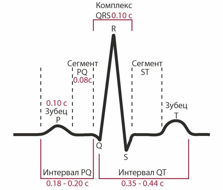 Characteristics of all indicators of an electrocardiographic study
 A prolonged PR interval (greater than 200 ms) may indicate first-degree heart block. Lengthening may be associated with hypokalemia, acute rheumatic fever or Lyme disease. A short PR interval (less than 120 ms) may be associated with Wolff-Parkinson-White syndrome or Lown-Ganong-Levine syndrome. PR segment depression may indicate atrial injury or pericarditis.
Examples of heart rhythm description and ECG interpretation
Normal sinus rhythm
Sinus rhythm is any heart rhythm in which the excitation of the heart muscle starts from the sinus node. It is characterized by correctly oriented P waves on the ECG. By convention, the term "normal" includes not only normal P waves, but all other ECG measurements.
 ECG norm and interpretation of all indicators
 ECG norm in adults:
- heart rate from 55 to 90 beats per minute;
- regular rhythm;
- normal PR interval, QT and QRS complex;
- The QRS complex is positive in almost all leads (I, II, AVF and V3-V6) and negative in aVR.
Sinus bradycardia
A heart rate less than 55 in sinus rhythm is called bradycardia. ECG decoding in adults should take into account all parameters: sports, smoking, medical history. Because in some cases, bradycardia is a variant of the norm, especially in athletes.
Pathological bradycardia occurs with weak sinus node syndrome and is recorded on the ECG at any time of the day. This condition is accompanied by constant fainting, pallor and hyperhidrosis. In extreme cases, with malignant bradycardia, pacemakers are prescribed.
 Sinus bradycardia
 Signs of pathological bradycardia:
- heart rate less than 55 beats per minute;
- sinus rhythm;
- P waves are vertical, consistent, and normal in morphology and duration;
- PR interval from 0.12 to 0.20 seconds;
Sinus tachycardia
The correct rhythm with a high heart rate (above 100 beats per minute) is called sinus tachycardia. Please note that normal heart rate varies with age, for example, in infants, the heart rate can reach 150 beats per minute, which is considered normal.
Advice! At home, with severe tachycardia, a strong cough or pressure on the eyeballs. These actions stimulate the vagus nerve, which activates the parasympathetic nervous system causing the heart to beat more slowly.
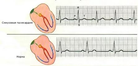 Sinus tachycardia
 Signs of pathological tachycardia:
- Heart rate above 100 beats per minute
- sinus rhythm;
- P waves are vertical, consistent and normal in morphology;
- the PR interval fluctuates between 0.12-0.20 seconds and decreases with increasing heart rate;
- QRS complex less than 0.12 seconds.
Atrial fibrillation
Atrial fibrillation is an abnormal heart rhythm characterized by rapid and irregular atrial contraction. Most episodes are asymptomatic. Sometimes an attack is accompanied by the following symptoms: tachycardia, fainting, dizziness, shortness of breath, or chest pain. The disease is associated with an increased risk of heart failure, dementia, and stroke.
 Atrial fibrillation
 Signs of atrial fibrillation:
- Heart rate unchanged or accelerated;
- P waves absent;
- electrical activity is chaotic;
- RR intervals are irregular;
- QRS complex less than 0.12 seconds (in rare cases, the QRS complex is lengthened).
Important! Despite the above explanations with data interpretation, the conclusion on the ECG should be made only by a qualified specialist - a cardiologist or general doctor. Deciphering the electrocardiogram and differential diagnosis requires higher medical education.
How to "read" myocardial infarction on an ECG?
For students beginning the study of cardiology, the question often arises, how to learn how to read a cardiogram correctly and identify myocardial infarction (MI)? You can “read” a heart attack on a paper tape by several signs:
- elevation of the ST segment;
- peaked T wave;
- deep Q wave or its absence.
In the analysis of the results of electrocardiography, these indicators are first of all identified, and then dealt with others. Sometimes the most early sign acute myocardial infarction is only a peaked T-wave. In practice, this is quite rare, because it appears only 3-28 minutes after the onset of a heart attack.
Peaked T-waves should be distinguished from peak T-waves associated with hyperkalemia. In the first few hours, the ST segments usually rise. Abnormal Q waves may appear within a few hours or after 24 hours.
An electrocardiogram is diagnostic method, allowing to determine the functional state the most important body the human body is the heart. Most people at least once in their lives have dealt with a similar procedure. But having received the result of the ECG, not every person, except perhaps with medical education, will be able to understand the terminology used in cardiograms.
What is cardiography
The essence of cardiography is the study of electrical currents that occur during the work of the heart muscle. The advantage of this method is its relative simplicity and availability. Strictly speaking, it is customary to call a cardiogram the result of measuring the electrical parameters of the heart, displayed in the form of a time graph.
The creation of electrocardiography in its modern form is associated with the name of the Dutch physiologist of the early 20th century, Willem Einthoven, who developed the basic methods of ECG and the terminology used by doctors to this day.
Thanks to the cardiogram, it is possible to obtain the following information about the heart muscle:
- Heart rate,
- Physical state of the heart
- The presence of arrhythmias
- The presence of acute or chronic damage to the myocardium,
- The presence of metabolic disorders in the heart muscle,
- The presence of violations of electrical conductivity,
- The position of the electrical axis of the heart.
Also, the electrocardiogram of the heart can be used to obtain information about some vascular diseases that are not related to the heart.
An ECG is usually performed in the following cases:
- Feeling an abnormal heartbeat;
- Attacks of shortness of breath, sudden weakness, fainting;
- Heartache;
- Murmurs in the heart;
- Deterioration of the condition of patients with cardiovascular diseases;
- Passage of medical examinations;
- Medical examination of people over 45 years old;
- Inspection before the operation.
- pregnancy;
- Endocrine pathologies;
- Nervous diseases;
- Changes in blood counts, especially with an increase in cholesterol;
- Over 40 years of age (once a year).
Where can I get a cardiogram?
If you suspect that your heart is not all right, then you can contact a general practitioner or cardiologist to give you a referral for an ECG. Also, on a paid basis, a cardiogram can be done at any clinic or hospital.
Procedure procedure
An EKG is usually taken in lying position. To take a cardiogram, a stationary or portable device is used - an electrocardiograph. Stationary devices are installed in medical institutions, and portable ones are used by emergency teams. The device receives information about the electrical potentials on the surface of the skin. To do this, electrodes are attached to the chest and limbs.
These electrodes are called leads. On the chest and limbs, 6 leads are usually installed. The chest leads are designated V1-V6, the leads on the limbs are called the main (I, II, III) and enhanced (aVL, aVR, aVF). All leads give a slightly different pattern of fluctuations, however, summing up the information from all the electrodes, you can find out the details of the work of the heart as a whole. Sometimes additional leads (D, A, I) are used.
Usually, the cardiogram is displayed as a graph on paper containing millimeter markings. Each lead-electrode has its own schedule. The standard belt speed is 5 cm/s, other speeds can be applied. In the cardiogram displayed on the tape, the main parameters, norm indicators and the conclusion generated automatically can also be indicated. Also, data can be recorded in memory and on electronic media.
After the procedure, it is usually required to decipher the cardiogram by an experienced cardiologist.
Holter monitoring
In addition to stationary devices, there are also portable devices for daily (Holter) monitoring. They are attached to the patient's body along with electrodes and record all the information received over a long period of time (usually within a day). This method gives much more complete information about the processes in the heart compared to a conventional cardiogram. So, for example, when taking a cardiogram in stationary conditions, the patient must be at rest. Meanwhile, some deviations from the norm can manifest themselves during physical exertion, in sleep, etc. Holter monitoring provides information about such phenomena.

Other types of procedures
There are several other methods of carrying out the procedure. For example, this is monitoring with physical activity. Abnormalities are usually more pronounced on the exercise ECG. The most common way to provide the body with the necessary physical activity is a treadmill. This method is useful in cases where pathologies can manifest themselves only in case of increased work of the heart, for example, if coronary disease is suspected.
With phonocardiography, not only the electrical potentials of the heart are recorded, but also the sounds that arise in the heart. The procedure is prescribed when it is necessary to clarify the occurrence of heart murmurs. This method often used for suspected heart defects.
The patient must be calm during the procedure. Between physical activity and the procedure must pass a certain period of time. It is also not recommended to undergo the procedure after eating, drinking alcohol, caffeinated drinks or cigarettes.
Causes that can affect the ECG:
- Times of Day,
- electromagnetic background,
- Physical exercise,
- food intake,
- The position of the electrodes.
Prong types
First, let's talk a little about how the heart works. It has 4 chambers - two atria and two ventricles (left and right). The electrical impulse, due to which it is reduced, is formed, as a rule, in the upper part of the myocardium - in the sinus pacemaker - the nerve sinoatrial (sinus) node. The impulse propagates down the heart, first affecting the atria and causing them to contract, then it passes through the atrioventricular ganglion and another ganglion - the bundle of His, and reaches the ventricles. The main burden of pumping blood is taken by the ventricles, especially the left one, which is involved in big circle circulation. This stage is called contraction of the heart or systole.
After the contraction of all parts of the heart, the time comes for their relaxation - diastole. Then the cycle repeats again and again - this process is called the heartbeat.
The state of the heart, in which there is no change in the propagation of impulses, is reflected on the ECG in the form of a straight horizontal line, called the isoline. The deviation of the graph from the isoline is called a tooth.
One heartbeat on an ECG contains six teeth: P, Q, R, S, T, U. The teeth can be directed both up and down. In the first case, they are considered positive, in the second - negative. The Q and S waves are always positive, and the R wave is always negative.

The teeth reflect the different phases of the contraction of the heart. P reflects the moment of contraction and relaxation of the atria, R - excitation of the ventricles, T - relaxation of the ventricles. Special designations are also used for segments (gaps between adjacent teeth) and intervals (sections of the graph, including segments and teeth), for example, PQ, QRST.
Correspondence of the stages of heart contraction and some elements of cardiograms:
- P - atrial contraction;
- PQ - horizontal line, the transition of the discharge from the atria through the atrioventricular node to the ventricles. The Q wave may be absent normally;
- QRS - ventricular complex, the element most often used in diagnostics;
- R - excitation of the ventricles;
- S - relaxation of the myocardium;
- T - relaxation of the ventricles;
- ST - horizontal line, myocardial recovery;
- U - may be absent in the norm. The reasons for the appearance of a tooth have not been clearly elucidated, however, the tooth is valuable for diagnosing certain diseases.
Below are some ECG abnormalities and their possible explanations. This information, of course, does not negate the fact that it is more expedient to entrust the decoding to a professional cardiologist who knows better all the nuances of deviations from the norms and related pathologies.
Major abnormalities and diagnosis
| Description | Diagnosis |
| The distance between the R teeth is not the same | atrial fibrillation, heart block, weakness of the sinus node, extrasystole |
| P wave too high (more than 5 mm), too wide (more than 5 mm), consists of two halves | atrial thickening |
| P wave absent in all leads except V1 | the rhythm does not come from the sinus node |
| PQ interval lengthened | atrioventricular block |
| QRS extension | ventricular hypertrophy, bundle branch block |
| No gaps between QRS | paroxysmal tachycardia, ventricular fibrillation |
| QRS in the form of a flag | heart attack |
| Deep and wide Q | heart attack |
| Wide R (more than 15 mm) in leads I, V5, V6 | left ventricular hypertrophy, bundle branch block |
| Deep S in III, V1, V2 | left ventricular hypertrophy |
| S-T above or below the isoline by more than 2 mm | ischemia or infarction |
| Tall, double-humped, pointed T | cardiac overload, ischemia |
| T merging with R | acute infarction |
Table of cardiogram parameters in adults
The norm of the duration of the elements of the cardiogram in children
The norms indicated in the table may also depend on age.
Rhythm of contractions
Violation of the rhythm of contractions is called. Rhythm irregularity in arrhythmia is measured as a percentage. O wrong rhythm indicates a deviation of the distance between similar teeth by more than 10%. Sinus arrhythmia, that is, arrhythmia associated with sinus rhythm, may be a variant of the norm for adolescents and young adults, but in most cases indicates the onset of a pathological process.
A kind of arrhythmia is extrasystole. He is told to her in the case when extraordinary reductions are observed. Single extrasystoles (no more than 200 per day with Holter monitoring) can also be observed in healthy people. Frequent extrasystoles that appear on the cardiogram in the amount of several pieces may indicate ischemia, myocarditis, heart defects.
Heart rate
This option is the most simple and clear. It determines the number of contractions in one minute. The number of contractions may be higher than normal (tachycardia) or lower than normal (bradycardia). The normal heart rate for adults can range from 60 to 80 beats. However, the norm in this case is a relative concept, so bradycardia and tachycardia may not always be evidence of pathology. Bradycardia can occur during sleep or in trained people, and tachycardia can occur during stress, after physical exertion, or at elevated temperature.
Heart rate norms for children of different ages

Photo: Africa Studio/Shutterstock.com
Types of heart rate
There are several types of heart rhythm, depending on where the nerve impulse begins to spread, leading to the contraction of the heart:
- sinus,
- atrial,
- atrioventricular,
- Ventricular.
Normally, the rhythm is always sinus. In this case, sinus rhythm can be combined with both a heart rate above normal and a heart rate below normal. All other types of rhythms are evidence of problems with the heart muscle.
atrial rhythm
Atrial rhythm also often appears on the cardiogram. Is the atrial rhythm normal or is it a kind of pathology? In most cases, the atrial rhythm on the ECG is not normal. However, this is a relatively mild degree of cardiac arrhythmias. It occurs in case of oppression or disruption of the sinus node. Possible reasons- ischemia, hypertension, sick sinus syndrome, endocrine disorders. However, individual episodes of atrial contractions can be observed in healthy people. This type rhythm can take on both the nature of bradycardia and the nature of tachycardia.
atrioventricular rhythm
Rhythm emanating from the atrioventricular node. In atrioventricular rhythm, the pulse rate usually drops to less than 60 beats per minute. Causes - weakness of the sinus node, atrioventricular blockade, taking certain drugs. Atrioventricular rhythm, combined with tachycardia, can occur during heart surgery, rheumatism, heart attack.
Ventricular rhythm
In ventricular rhythm, contractile impulses propagate from the ventricles. The contraction rate drops below 40 beats per minute. The most severe form of rhythm disturbance. Occurs in acute myocardial infarction, heart defects, cardiosclerosis, insufficiency cardiac circulation, in the preagonal state.
Electrical axis of the heart
Another important parameter is the electrical axis of the heart. It is measured in degrees and reflects the direction of propagation of electrical impulses. Normally, it should be somewhat inclined to the vertical and be 30-69º. At an angle of 0-30º, they talk about the horizontal location of the axis, at an angle of 70-90º - about the vertical. Axial deviation in one direction or another may indicate a disease, for example, hypertension or intracardiac blockades.
What do the conclusions on cardiograms mean?
Consider some of the terms that an ECG decoding may contain. They do not always indicate serious pathologies, however, in any case, they require a visit to a doctor for advice, and sometimes additional examinations.

Photo: Have a nice day Photo/Shutterstock.com
Atrioventricular block
It is reflected on the graph as an increase in the duration of the P-Q interval. 1 degree of the disease is reflected in the form of a simple prolongation of the interval. Grade 2 is accompanied by a deviation of the QRS parameters (loss of this complex). At grade 3, there is no connection between P and the ventricular complex, which means that the ventricles and atria work each in their own rhythm. The syndrome in stages 1 and 2 is not life-threatening, but requires treatment, as it can go into an extremely dangerous stage 3, in which the risk of cardiac arrest is high.
Ectopic rhythm
Any heart rhythm other than sinus. May indicate the presence of blockades, coronary heart disease, or be a variant of the norm. It can also appear as a result of an overdose of glycosides, neurocirculatory dystonia, hypertension.
Sinus bradycardia or tachycardia
Sinus rhythm on the ECG that is below (bradycardia) or above (tachycardia) the normal range. It can be both a variant of the norm and be a symptom of some pathologies. However, in the latter case, this symptom will most likely not be the only one indicated in the transcript of the cardiogram.
Nonspecific ST-T wave changes
What it is? This entry suggests that the reasons for the interval change are unclear and more research is needed. May indicate a violation metabolic processes in the body, for example, a change in the balance of potassium, magnesium, sodium ions, or endocrine disorders.
Conduction disorders within the ventricles
As a rule, they are associated with conduction disturbances inside the His nerve bundle. May affect the trunk of the beam or its legs. May lead to delayed contraction of one of the ventricles. Direct therapy for blockades of the His bundle is not carried out, only the disease that caused them is treated.
Incomplete blockade of the right leg of the bundle of His (RBBBB)
Widespread violation of ventricular conduction. In most cases, however, it does not lead to the development of pathologies and is not their consequence. If the patient does not have problems with the cardiovascular system, then this symptom does not require treatment.
Complete right bundle branch block (RBBBB)
This violation is more serious than an incomplete blockade. May indicate myocardial damage. It usually occurs in older and older people, it is rarely found in children and adolescents. Possible symptoms- shortness of breath, dizziness, general weakness and fatigue.
Blockade of the anterior branch of the left leg of the bundle of His (BPVLNPG)
Occurs in patients with hypertension who have had a heart attack. It may also indicate cardiomyopathy, cardiosclerosis, atrial septal defect, mitral valve insufficiency. Has no characteristic symptoms. It is observed mainly in the elderly (over 55 years).
Blockade of the posterior branch of the left leg of the bundle of His (B3VLNPG)
As a separate symptom, it is rare, as a rule, it is combined with blockade of the right bundle leg. May indicate a heart attack, cardiosclerosis, cardiomyopathy, calcification of the conduction system. The blockade is indicated by a deviation in the electrical axis of the heart to the right.
Metabolic changes
Reflect malnutrition of the heart muscle. First of all, it concerns the balance of potassium, magnesium, and sodium. The syndrome is not an independent disease, but indicates other pathologies. It can be observed with ischemia, cardiomyopathy, hypertension, rheumatism, cardiosclerosis.
Low voltage ECG
Electrodes installed on the patient's body capture currents of a certain voltage. If the voltage parameters are below normal, then they talk about low voltage. This indicates insufficient external electrical activity of the heart and may be the result of pericarditis or a number of other diseases.
Paroxysmal tachycardia
A rare condition that differs from normal (sinus) tachycardia, primarily in that it has a very high heart rate - more than 130 beats / s. In addition, the basis of paroxysmal tachycardia is the incorrect circulation of an electrical impulse in the heart.
Atrial fibrillation
At the core atrial fibrillation are atrial fibrillation or flutter. Arrhythmia caused by atrial fibrillation can also occur in the absence of heart pathologies, for example, with diabetes, intoxication, and also with smoking. Atrial flutter may be characteristic of cardiosclerosis, some types of coronary disease, inflammatory processes myocardium.
Sinoatrial blockade
Difficulty in the output of the impulse from the sinus (sinoatrial) node. This syndrome is a type of sick sinus syndrome. It is rare, mostly in the elderly. Possible causes are rheumatism, cardiosclerosis, calcification, severe hypertension. May lead to severe bradycardia, syncope, convulsions, respiratory failure.
Hypertrophic conditions of the myocardium
They indicate an overload of certain parts of the heart. The body feels this situation and reacts to it by thickening the muscular walls of the corresponding department. In some cases, the causes of the condition may be hereditary.
Myocardial hypertrophy
General myocardial hypertrophy is a protective reaction indicating excessive load on the heart. May lead to arrhythmia or heart failure. Sometimes it is the result of a heart attack. A variation of the disease is hypertrophic cardiomyopathy - a hereditary disease that leads to an abnormal arrangement of heart fibers and carries a risk of sudden cardiac arrest.
Left ventricular hypertrophy
The most common symptom, which does not always indicate severe pathologies of the heart. May be characteristic of arterial hypertension, obesity, some heart defects. Sometimes it is also observed in trained people, people engaged in heavy physical labor.
Right ventricular hypertrophy
A rarer, but at the same time much more dangerous symptom than left ventricular hypertrophy. Indicates insufficiency of pulmonary circulation, severe lung diseases, valvular defects or severe heart defects (tetralogy of Fallot, ventricular septal defect).
Left atrial hypertrophy
It is reflected in the form of a change in the P wave on the cardiogram. With this symptom, the tooth has a double top. Indicates mitral or aortic stenosis, hypertension, myocarditis, cardiomyopathies. Leads to chest pain, shortness of breath, fatigue, arrhythmias, fainting.
Right atrial hypertrophy
Less common than left atrial hypertrophy. It can have many causes - pulmonary pathologies, chronic bronchitis, arterial embolism, tricuspid valve defects. Sometimes seen during pregnancy. May lead to circulatory disorders, edema, shortness of breath.
Normocardia
Normocardia or normosystole means normal frequency heart contractions. However, the presence of normosystole in itself is not evidence that the ECG is normal and everything is in order with the heart, since it may not exclude other pathologies, such as arrhythmias, conduction disorders, etc.
Nonspecific T wave changes
This symptom is typical for about 1% of people. A similar conclusion is made if it cannot be unambiguously associated with any other disease. Thus, with non-specific changes in the T wave, additional studies are needed. The symptom may be characteristic of hypertension, ischemia, anemia and some other diseases, and may also occur in healthy people.
tachysystole
Also often called tachycardia. This is the general name for a number of syndromes in which there is an increased frequency of contractions of various parts of the heart. There are ventricular, atrial, supraventricular tachysystoles. Such types of arrhythmias as paroxysmal tachycardia, atrial fibrillation and flutter also belong to tachysystoles. In most cases, tachysystoles are a dangerous symptom and require serious treatment.
ST depression of the heart
ST segment depression is common in high-frequency tachycardias. Often it indicates a lack of oxygen supply to the heart muscle and may be characteristic of coronary atherosclerosis. At the same time, the appearance of depression in healthy people is also noted.
Borderline ECG
This conclusion often frightens some patients who have found it on their cardiograms and are inclined to think that "marginal" means almost "mortal". In fact, such a conclusion is never given by a doctor, but is generated by a program that analyzes the parameters of the cardiogram on an automatic basis. Its meaning is that a number of parameters go beyond the norm, but it is impossible to unequivocally conclude that there is any pathology. Thus, the cardiogram is on the border between normal and pathological. Therefore, upon receipt of such a conclusion, a doctor's consultation is required, and, perhaps, everything is not so scary.
Pathological ECG
What it is? This is a cardiogram, which clearly showed some serious deviations from the norm. These can be arrhythmias, conduction disorders or nutrition of the heart muscle. Pathological changes require immediate consultation with a cardiologist, who must indicate the treatment strategy.
Ischemic changes on the ECG
Ischemic disease is caused by impaired blood circulation in the coronary vessels of the heart and can lead to such serious consequences as myocardial infarction. Therefore, the identification of ischemic signs on the ECG is a very important task. Ischemia on early stage can be diagnosed by changes in the T wave (rise or fall). With more late stage there are changes in the ST segment, and in acute - changes in the Q wave.
Deciphering the ECG in children
In most cases, deciphering the cardiogram in children is simple. But the parameters of the norm and the nature of the violations may differ compared to those in adults. So, children normally have a much more frequent heartbeat. In addition, the sizes of teeth, intervals and segments are somewhat different.


